Background
Alkaline phosphatase (AP), most commonly sourced from calf intestine (CIAP) or produced recombinantly, is the second most widely deployed reporter enzyme in immunoassay diagnostics after HRP. While HRP dominates rapid, high-throughput screening applications, AP occupies a distinct and indispensable niche where signal stability, low endogenous background, and broad dynamic range are paramount. Its larger molecular size (ca. 140 kDa dimer for mammalian AP) and slower catalytic turnover relative to HRP result in signal accumulation rather than rapid burst kinetics—an advantage in quantitative assays that demand extended incubation and precise low-end discrimination.
However, AP's dependence on zinc and magnesium cofactors, sensitivity to chelating agents, and propensity for inactivation under suboptimal storage conditions make conjugate design more demanding than HRP labeling. Creative Enzymes Diagnostic offers a specialized AP Conjugation Service that addresses these challenges through gentle coupling chemistries, metal-ion-preserving formulations, and rigorous activity validation to deliver conjugates optimized for high-sensitivity diagnostic platforms.
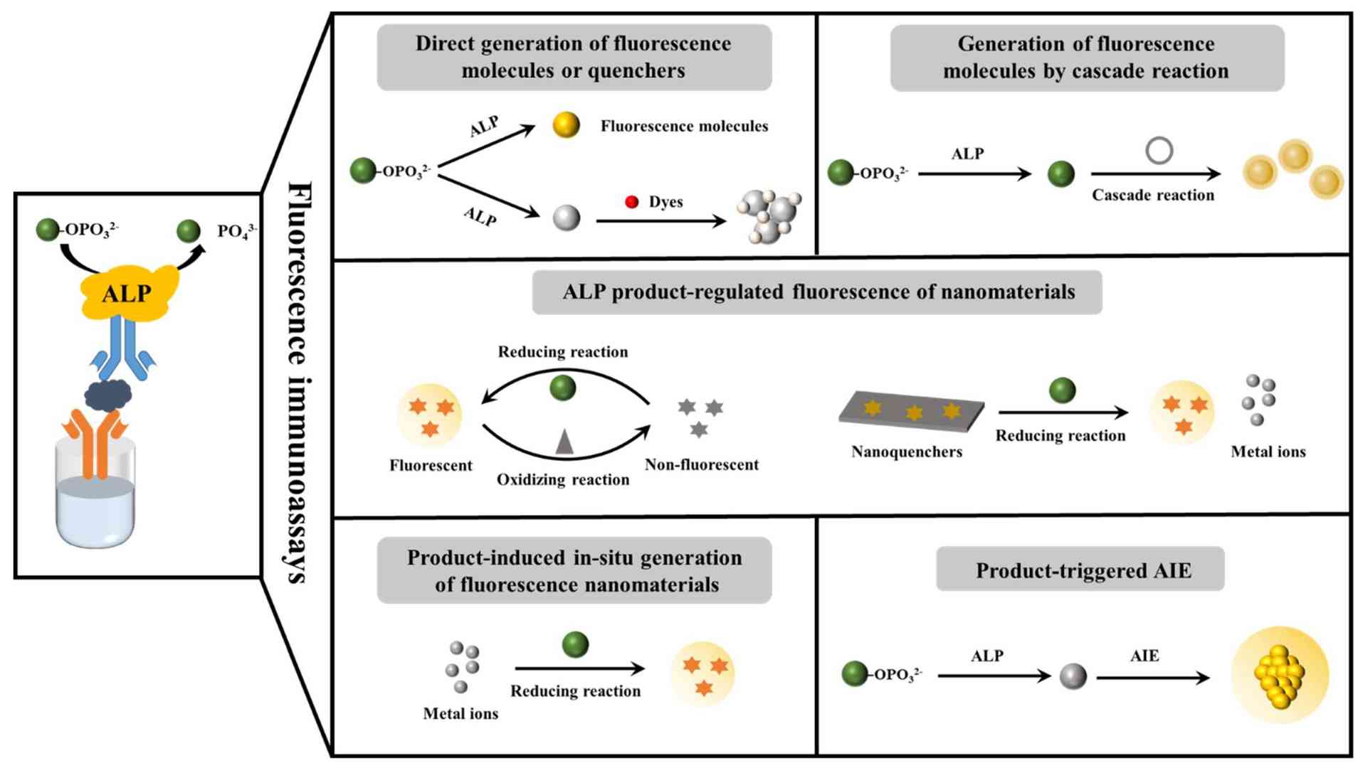 Figure 1. Schematic diagram outlining the alkaline-phosphatase-linked fluorescence immunoassays. (Li et al., 2023)
Why Choose AP
AP is not merely an alternative to HRP—it offers fundamentally different kinetic and biochemical properties that make it the superior choice for specific assay architectures. Understanding these distinctions ensures optimal reporter selection during assay development.
High Signal Stability
- AP catalyzes substrate dephosphorylation with slower turnover kinetics than HRP, producing a steady, linear signal accumulation over extended incubation periods rather than the rapid burst-and-decay profile characteristic of peroxidase reactions
- Prolonged signal integration enables higher sensitivity in endpoint assays by allowing incubation times of 30–60 minutes (or longer) without significant substrate depletion or signal self-quenching, which commonly limits HRP-based detection at low analyte concentrations
- Reduced enzyme inactivation during the assay window because AP does not generate reactive oxygen species (H2O2-derived radicals) that can damage the conjugate or adjacent assay components, a known limitation of HRP catalysis
Low Endogenous Interference
- Mammalian tissues and serum samples contain relatively low levels of endogenous AP activity compared to the ubiquitous presence of endogenous peroxidases in erythrocytes, leukocytes, and plant-derived food matrices that plague HRP-based IHC and serum ELISA
- Minimal background in blood-based diagnostics because hemoglobin-associated peroxidase activity— a major confounder in HRP assays—is entirely absent; AP conjugates therefore require less aggressive blocking and sample pre-treatment
- Strategic advantage in tissue-based assays (IHC, ICC) where endogenous peroxidase activity in myeloid cells and red blood cells necessitates methanol/H2O2 quenching that can damage epitopes; AP detection avoids this destructive pre-treatment
Excellent Dynamic Range
- AP-mediated signal generation exhibits a broader linear response range across analyte concentrations, reducing hook effects and enabling accurate quantification across three to four orders of magnitude without dilution and re-assay
- Superior performance in competitive immunoassays and low-abundance biomarker detection where the extended linearity of AP chemiluminescence substrates (e.g., CSPD, CDP-Star) delivers lower limits of detection (LOD) than equivalent HRP systems
Multiple Substrate Options
- Chromogenic substrates including p-nitrophenyl phosphate (pNPP) for soluble yellow endpoint detection in ELISA, and 5-bromo-4-chloro-3-indolyl phosphate (BCIP) combined with nitroblue tetrazolium (NBT) for insoluble purple precipitate formation in Western blot and IHC
- Chemiluminescent substrates such as CSPD and CDP-Star (1,2-dioxetane-based substrates) that generate sustained glow-type luminescence with femtogram-level detection sensitivity and extended emission half-lives ideal for automated luminometer integration
- Fluorogenic substrates including 4-methylumbelliferyl phosphate (4-MUP), which yields highly fluorescent 4-methylumbelliferone upon dephosphorylation, enabling multiplexed fluorescence detection alongside other fluorophore channels
AP Conjugation Technologies
AP's complex quaternary structure and cofactor requirements demand coupling chemistries that preserve enzymatic integrity. We employ methods that minimize disruption to the active site metal center and maintain subunit association.
Covalent Coupling
- Glutaraldehyde-mediated two-step conjugation for antibody-AP linkage, utilizing the high density of surface lysines on both proteins; reaction conditions are carefully titrated to prevent intra-AP cross-linking that would inactivate the dimeric enzyme
- Heterobifunctional cross-linkers (Sulfo-SMCC, GMBS) that form stable thioether bonds between thiol-activated AP and maleimide-derivatized antibodies, offering greater control over conjugation geometry than homobifunctional reagents
- NHS-ester/EDC chemistry for zero-length coupling of carboxyl-rich AP surfaces to antibody amines, performed in metal-ion-supplemented buffers (Zn2+, Mg2+) to prevent cofactor stripping and irreversible apoenzyme formation
Site-Specific Labeling
- Carbohydrate-directed conjugation exploiting the N-linked glycosylation sites on mammalian AP; mild periodate oxidation of glycan chains generates aldehydes for reductive amination to antibody lysines, directing coupling away from the catalytic center
- Engineered cysteine handles introduced into the antibody Fc region or AP C-terminus via recombinant expression, enabling directional thiol-maleimide coupling that preserves both the antigen-binding paratope and the AP active site
- Enzymatic ligation using sortase A or microbial transglutaminase to attach AP to recognition-tagged antibodies under mild, physiological conditions that eliminate harsh chemical cross-linkers and preserve native protein folding
Controlled Stoichiometry
- Systematic variation of AP-to-antibody molar ratios (typically 1:1 to 3:1 for random coupling) to identify the optimal degree of labeling that maximizes signal output while preventing antibody cross-linking and AP dimer disruption
- Analytical SEC fractionation to isolate monodisperse 1:1 conjugate species from free AP, free antibody, and higher-order aggregates, with real-time UV/Vis monitoring at 278 nm and 405 nm (AP absorbance maximum)
- Validation that controlled stoichiometry reduces batch variability; site-specific 1:1 conjugates exhibit coefficient of variation (CV)
Performance Optimization
AP conjugate performance depends critically on preserving cofactor integrity, minimizing non-specific binding, and developing storage formulations that prevent metal loss and subunit dissociation. Our optimization workflow targets each vulnerability.
Enzyme Activity
- Pre-conjugation AP activity verification using pNPP hydrolysis assay (405 nm) to establish baseline specific activity; only AP lots with specific activity > 3,000 U/mg are qualified for conjugation
- Post-conjugation kinetic characterization (kcat, KM) with pNPP and chemiluminescent substrates to confirm that coupling chemistry has not disrupted the binuclear metal center (Zn2+, Mg2+) or allosteric subunit communication
- Metal-ion supplementation (1 mM MgCl2, 0.1 mM ZnCl2) in all processing buffers to prevent formation of catalytically inactive apo-AP during purification and storage
Signal-to-Noise Ratio
- Optimization of blocking reagents (casein, fish gelatin, proprietary synthetic blockers) to suppress non-specific adsorption of the large, charged AP conjugate to polystyrene microplate surfaces and PVDF/nitrocellulose membranes
- Titration of conjugate working concentration against zero-calibrator wells to identify the dilution that yields maximal specific signal with minimal background, typically 10- to 100-fold lower concentration than equivalent HRP conjugates due to AP's sustained signal generation
- Assessment of cross-reactivity with unrelated assay components and sample matrix interference (hemolysis, lipemia, icterus) to validate assay robustness in clinical specimen types
Long-Term Stability
- Accelerated degradation studies at 37°C with weekly activity measurements to project real-time shelf-life; Arrhenius modeling predicts 12-month stability at 4°C and 24-month stability at −20°C for optimized formulations
- Freeze-thaw cycle testing (10 cycles, −80°C to 25°C) to identify variants and formulations resistant to ice-crystal damage and metal-ion precipitation that commonly inactivate AP
- Monitoring of aggregation propensity by dynamic light scattering (DLS) and turbidity measurement; batches with polydispersity index > 0.15 are rejected to prevent assay background drift over storage
Storage Buffer Development
- Formulation screening with stabilizers (trehalose, sucrose, BSA, glycerol) and divalent cation chelators (EDTA avoidance) to identify buffers that maintain AP holoenzyme integrity and prevent subunit dissociation
- pH optimization (typically pH 7.4–8.0) to balance AP structural stability against the risk of alkaline denaturation; Tris-HCl and diethanolamine buffers evaluated for compatibility with intended assay conditions
- Preservative evaluation (sodium azide, ProClin, gentamicin) to ensure antimicrobial protection without AP inhibition, as some preservatives can chelate metal ions or interfere with the active site nucleophile
Typical Applications
AP conjugates excel in assay formats where signal linearity, low background, and extended incubation windows provide analytical advantages over peroxidase-based detection.
- ELISA: High-sensitivity sandwich and competitive ELISAs for cytokines, hormones, and infectious disease markers where wide dynamic range and low-end precision are critical. AP-pNPP endpoint assays offer excellent reproducibility in manual and semi-automated workflows, while AP chemiluminescence substrates extend sensitivity into the sub-picogram-per-milliliter range for troponin, PTH, and other low-abundance analytes.
- CLIA: Automated chemiluminescence immunoassay platforms utilizing dioxetane substrates (CSPD, CDP-Star) with glow-kinetics emission profiles that integrate reliably over 5–30 minute read windows. AP's lack of H2O2 dependence eliminates reagent instability and instrument maintenance issues associated with peroxidase-based CLIA systems.
- Microarray: Protein and antibody microarrays for biomarker discovery and validation where AP-mediated tyramide signal amplification (TSA) or direct fluorescent substrate conversion enables high-density, multiplexed detection. AP's signal stability supports the extended scanning and image acquisition times required for high-resolution microarray readers without significant signal decay between slides.
FAQs
Q1. When should I choose AP over HRP for my immunoassay?
A1. Choose AP when your assay requires (1) extended signal accumulation for maximum sensitivity, (2) low background in samples with high endogenous peroxidase activity (blood, tissue), (3) a broad dynamic range without dilution, or (4) compatibility with phosphate-buffered assay conditions that inhibit HRP. HRP remains preferable for rapid, high-throughput assays and applications requiring small conjugate size. We can evaluate both reporters in parallel during feasibility to guide selection.
Q2. Does AP conjugation require special handling compared to HRP?
A2. Yes. AP requires divalent metal cofactors (Zn2+ and Mg2+) for catalytic activity, so all conjugation buffers must be supplemented with these ions and free of metal chelators like EDTA or EGTA. AP is also more sensitive to freeze-thaw cycles and alkaline pH extremes. Our protocols use gentle cross-linking conditions, metal-supplemented buffers, and cryoprotectant formulations specifically designed to preserve AP holoenzyme integrity throughout manufacturing and storage.
Q3. What substrates do you validate for your AP conjugates?
A3. We validate all conjugates with the three major substrate classes: chromogenic (pNPP for ELISA; BCIP/NBT for membrane and tissue applications), chemiluminescent (CSPD and CDP-Star for high-sensitivity CLIA), and fluorogenic (4-MUP for fluorescence microplate and microarray detection). We can additionally validate performance with client-specified proprietary substrates or substrate cocktails upon request.
Q4. What is the typical degree of labeling for AP-antibody conjugates?
A4. Because AP is significantly larger than HRP (~140 kDa vs. ~44 kDa), we typically target a lower degree of labeling (DOL) of 1–2 AP molecules per IgG antibody to prevent steric hindrance of antigen binding and aggregation. Site-specific conjugates are manufactured at a defined 1:1 ratio. The optimal DOL is confirmed by UV/Vis spectrophotometry (A405/A280 ratio) and validated functionally in the target assay format.
Q5. Can AP conjugates be lyophilized for ambient-temperature distribution?
A5. Yes, but lyophilization requires specialized formulation development to prevent metal-ion loss and subunit dissociation during drying and reconstitution. We have developed proprietary stabilizer matrices containing trehalose, metal salts, and compatible buffers that maintain >90% AP activity after lyophilization and 6-month ambient storage. Lyophilized AP conjugates are particularly valuable for point-of-care diagnostics and resource-limited settings.
Q6. What is the typical timeline and deliverable for an AP conjugation project?
A6. A standard custom AP conjugation project spans 6–8 weeks, including 1–2 weeks for antibody feasibility and conjugation chemistry selection, 2–3 weeks for conjugation and purification, and 2–3 weeks for analytical characterization and assay validation. Deliverables include the purified conjugate, a Certificate of Analysis (CoA) with activity, binding, DOL, and purity data, and a stability report. Expedited 4-week timelines are available for urgent diagnostic development programs.
Creative Enzymes Diagnostic leverages specialized expertise in metal-dependent enzyme biochemistry and advanced conjugation chemistry to deliver AP conjugates that outperform standard peroxidase systems in demanding quantitative and high-sensitivity applications. From ELISA development to automated CLIA and multiplexed microarray platforms, our AP Conjugation Service provides the signal stability, low background, and batch consistency your diagnostic assay requires.
Contact our business development team today to discuss your AP conjugation requirements!
References
- Liu L, Chang Y, Lou J, Zhang S, Yi X. Overview on the development of alkaline-phosphatase-linked optical immunoassays. Molecules. 2023;28(18):6565. doi:10.3390/molecules28186565
Related Services

