Background
Lateral flow assays (LFAs) have become the backbone of point-of-care diagnostics, delivering rapid, instrument-free results in infectious disease screening, cardiac marker detection, and home testing. While colloidal gold dominates the visual readout market, enzyme-mediated signal generation offers unmatched sensitivity and quantitative potential for next-generation rapid tests. However, translating enzyme-substrate chemistry from the controlled environment of a microplate to the porous architecture of a nitrocellulose membrane introduces unique challenges: uneven flow dynamics, membrane-induced protein denaturation, substrate precipitation artifacts, and the need for dry-reagent stability under ambient storage. Creative Enzymes Diagnostic provides specialized enzyme signal development and optimization services tailored specifically for lateral flow and membrane-based diagnostic platforms, bridging the gap between classical enzyme immunoassay performance and the operational constraints of rapid test strips.
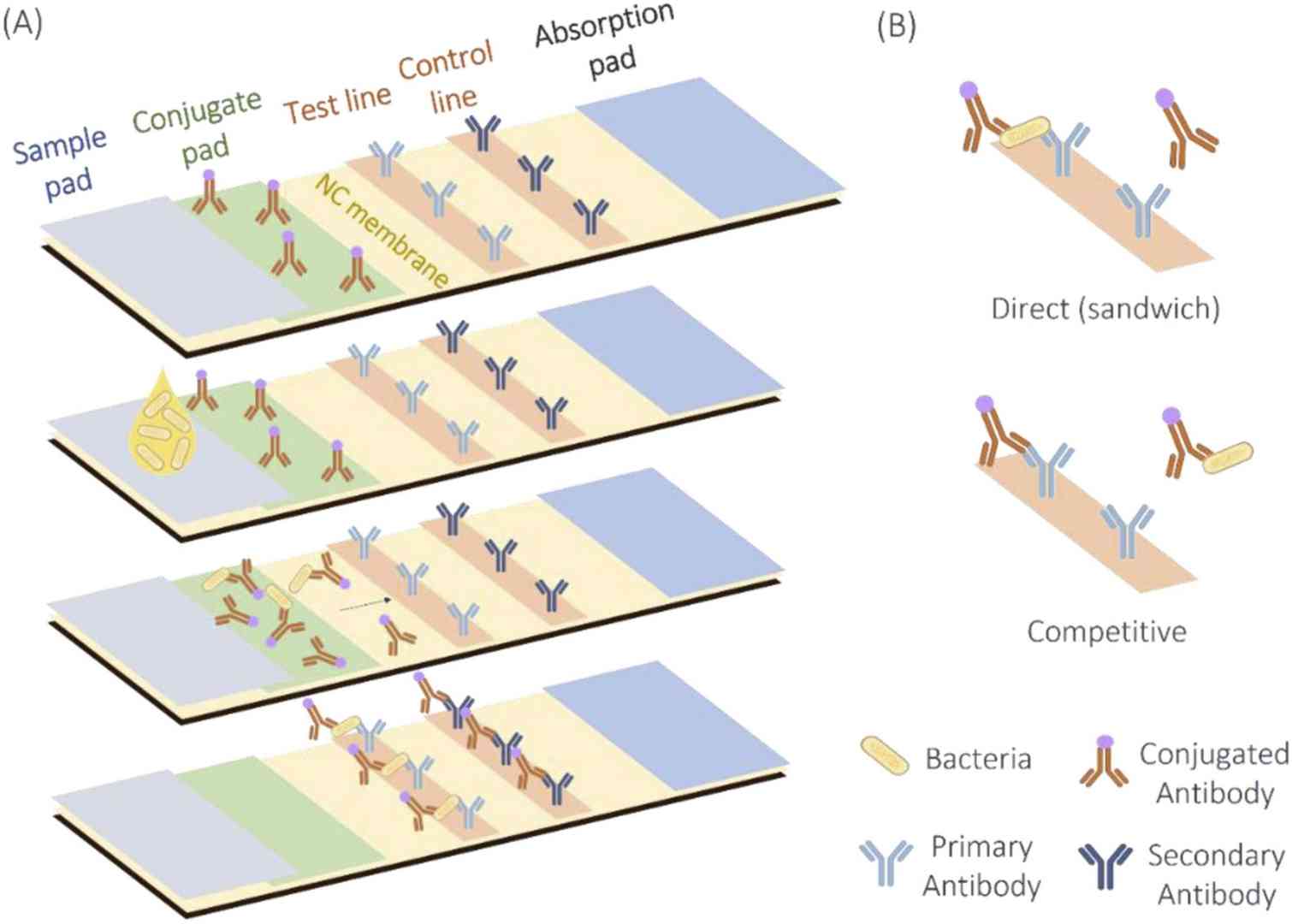 Figure 1. Lateral Flow Assay: (A) Basic structure and functioning of a direct LFA; (B) Direct and competitive types of LFA. (Santos et al., 2022)
Signal Development for Lateral Flow
Successful enzyme signal generation on membrane platforms requires rethinking every element of the signal cascade—from enzyme selection through substrate chemistry—to accommodate the porous matrix, capillary flow kinetics, and visual interpretation requirements of rapid tests.
Reporter Enzyme Selection
- Horseradish peroxidase (HRP) selected for its small size (~44 kDa), high turnover, and compatibility with insoluble precipitating substrates (4-CN, DAB, TMB precipitate) that generate sharp, localized color lines on nitrocellulose without excessive lateral diffusion; poly-HRP constructs evaluated for signal amplification in low-abundance analyte detection → HRP Conjugation Service for Immunoassays
- Alkaline phosphatase (AP) chosen for broad dynamic range and compatibility with BCIP/NBT and Fast Red substrates that produce stable, permanent precipitates resistant to fading under ambient light; AP's slower kinetics allow extended development times (5–15 minutes) that improve visual threshold detection compared to rapid gold-label readouts → Alkaline Phosphatase (AP) Conjugation Service
- β-galactosidase deployed for ultra-sensitive fluorescence or chemiluminescent readouts in reader-based quantitative LFAs, where its high molecular weight is offset by superior substrate turnover and minimal endogenous background in clinical matrices → β-Galactosidase, Urease and Oxidase Labeling Services
- Enzyme stability under dry-state storage prioritized; enzymes qualified by accelerated stress testing (37°C, 45°C) on conjugate pads to ensure >90% activity retention over the claimed shelf-life
Membrane Compatibility
- Nitrocellulose membrane pore size (typically 8–15 μm for standard LFAs, 5–10 μm for slower flow, higher sensitivity assays) optimized to balance capillary flow rate against protein binding capacity and line sharpness; membranes with intrinsic high protein binding (≥100 μg/cm2 BSA) preferred to immobilize capture antibodies without additional surface activation
- Membrane backing and plastic housing materials evaluated for compatibility with organic solvent-based substrate cocktails (DMSO, ethanol carriers) and acidic stop solutions; polyester or vinyl backing selected to prevent delamination during prolonged incubation with aqueous or alcoholic reagents
- Conjugate pad material (glass fiber, cellulose, rayon) screened for low intrinsic peroxidase activity (critical for HRP systems) and minimal pH shift upon rehydration; pad pretreatment with stabilizers (sucrose, trehalose, PVP) to preserve enzyme activity during drying and storage
- Wicking pad capacity calibrated to ensure complete sample and wash buffer traversal without premature flow stoppage, which can cause uneven substrate deposition and false line intensity variation
Running Buffer Optimization
- Surfactant selection (Tween-20, Triton X-100, SDS at 0.05–0.5% w/v) to control flow uniformity and reduce non-specific binding without inhibiting enzyme activity; excessive surfactant can strip enzyme-conjugate from the capture line or cause line spreading
- pH and ionic strength tuned to maintain enzyme activity while promoting antigen-antibody binding; phosphate-buffered saline (PBS, pH 7.4) standard for HRP systems, while AP-based LFAs require Tris or diethanolamine buffers at pH 8.5–9.5, necessitating either a two-buffer architecture or pH-adjusted substrate pads
- Blocking agents (BSA, casein, PVP, PEG) incorporated into the conjugate release pad and sample pad to suppress non-specific protein adsorption to the membrane matrix; fish gelatin and synthetic blockers evaluated for reduced cross-reactivity with heterophilic antibodies in serum and whole blood samples
- Viscosity modifiers (sucrose, trehalose, Ficoll) added to control flow rate and extend incubation time at the capture zone, improving binding efficiency without requiring manual timing steps
Signal Enhancement
- Precipitating substrate cocktails formulated for sharp line definition and high contrast; HRP systems optimized with 4-chloro-1-naphthol (4-CN) or DAB for dark purple/brown lines, while AP systems use BCIP/NBT for permanent blue-purple precipitates visible to the naked eye at picogram-per-milliliter analyte levels
- Signal amplification cascades including biotin-streptavidin-HRP layering, poly-HRP dendrimers, and HRP-urease coupled pH-shift indicators to boost signal intensity 10- to 50-fold over standard single-enzyme labeling, enabling visual detection thresholds approaching those of instrument-read fluorescence assays
- Silver enhancement for HRP-driven deposition of metallic silver onto gold nanoparticle seeds, creating black lines with extreme visual contrast; silver enhancer formulations optimized for uniform deposition without background grain formation on the membrane
- Substrate pad architecture designed as a separate, rehydratable pad rather than direct liquid addition, allowing controlled release of substrate solution across the capture zone and preventing flooding that causes line smearing
Assay Optimization
Membrane-based enzyme assays require optimization parameters distinct from microplate ELISA. We address sensitivity, flow kinetics, background, and stability as interdependent system variables.
Detection Sensitivity
- Enzyme conjugate loading on the release pad titrated to deliver sufficient signal at the cutoff concentration without causing prozone effects or hook phenomena in strongly positive samples
- Capture antibody line density optimized (typically 0.5–2.0 mg/mL dispensed at 1.0 μL/cm) to maximize antigen binding capacity while minimizing non-specific protein adsorption that elevates background
- Signal-to-cutoff (S/CO) ratio targeting >3.0 at the claimed detection limit for visual assays, and >10 for reader-based quantitative systems, with rigorous validation using clinical specimen panels near the threshold
Flow Performance
- Capillary flow time standardized (typically 60–180 seconds for a 25 mm membrane strip) to ensure complete sample traversal while allowing adequate residence time at the capture zone for antigen-antibody binding equilibrium
- Sample pad pretreatment with red blood cell separation agents (buffered saline, aggregated IgG) for whole blood applications, preventing hemoglobin interference with HRP-based color development
- Conjugate release kinetics synchronized with sample front migration; enzyme conjugate dried in a sugar matrix that dissolves within 5–10 seconds of sample contact to ensure homogeneous mixing before reaching the test line
Background Reduction
- Membrane blocking with protein and polymer cocktails after capture line dispensing to occupy non-specific binding sites without obscuring the test and control lines
- Enzyme quenching protocols for endogenous peroxidase in blood samples (sodium azide, methanol pretreatment of sample pad) validated for HRP-system compatibility; AP systems benefit from inherent low endogenous background in mammalian matrices
- Substrate blank optimization to eliminate membrane auto-coloration from substrate components; lot qualification of nitrocellulose membrane for intrinsic absorbance and fluorescence background
Shelf-Life Improvement
- Lyophilization or spray-drying of enzyme conjugates onto glass-fiber pads with trehalose/sucrose matrices, demonstrating >90% activity retention after 24 months at ambient temperature in foil pouches with desiccant
- Substrate pad stabilization through encapsulation in water-soluble films or separation from the enzyme by a physical barrier that dissolves upon sample addition, preventing premature enzyme-substrate contact during storage
- Accelerated aging studies (ICH Q1A(R2)) at 37°C and 45°C with Arrhenius modeling to establish expiration dating and support regulatory claims for room-temperature distribution in resource-limited settings
Compatible Platforms
Our enzyme signal systems are adapted to the structural and operational requirements of each membrane-based platform, from standard dipstick formats to multiplexed microfluidic paper devices.
| Platform |
Architecture & Flow Characteristics |
Enzyme Signal Approach |
Typical Applications |
| Lateral Flow Assays |
Standard dipstick format with sample pad, conjugate pad, nitrocellulose membrane, and wicking pad; capillary flow driven by absorbent wick |
Precipitating substrates (DAB, BCIP/NBT, 4-CN) for visual line readout; poly-HRP amplification for enhanced sensitivity; quantitative reader compatibility with reflectance measurement |
Infectious disease (HIV, influenza, malaria), cardiac markers (troponin, NT-proBNP), fertility (hCG, LH), drugs of abuse |
| Nitrocellulose Membrane Assays |
Dot blot, slot blot, or line blot formats without integrated flow path; manual sample application and wash steps |
Direct membrane enzyme immunoassay with precipitating or soluble substrates; multiple antigen dots per membrane for parallel screening; stripping and reprobing protocols for HRP systems |
Autoantibody panels, allergen screening, serological profiling, research protein detection |
| Paper-Based Diagnostics |
Microfluidic paper analytical devices (μPADs) with hydrophobic wax-printed channels; cellulose fiber matrix |
Colorimetric enzyme reactions (HRP-TMB, AP-pNPP) in confined channels; distance-based or intensity-based quantification without instrumentation; glucose oxidase for self-powered assays |
Glucose monitoring, water quality testing, environmental contaminant detection, resource-limited POC settings |
| Multiplex Rapid Tests |
Single strip with multiple parallel test lines or arrayed capture zones; sample split across independent channels |
Multi-enzyme labeling (HRP + AP) with spectrally distinct precipitates (brown + blue) for visual differentiation; fluorescence enzyme substrates for reader-based multiplex quantification |
Respiratory pathogen panels (COVID-19/flu A/B), sexually transmitted infection combos, tropical disease multiplex screening |
Our Workflow
Every lateral flow enzyme signal project follows a structured development pathway from feasibility through manufacturing transfer, ensuring that the final product meets performance, stability, and regulatory requirements.

FAQs
Q1. Why would I choose an enzyme label over colloidal gold for my lateral flow assay?
A1. Enzyme labels offer 10- to 100-fold higher sensitivity than standard 40 nm gold nanoparticles due to catalytic signal amplification—each enzyme molecule generates thousands of detectable product molecules. Enzymes also enable quantitative reader-based detection with broader dynamic ranges, and precipitating substrates produce permanent, non-fading records. Gold remains advantageous for simple, low-cost visual assays where moderate sensitivity is sufficient.
Q2. Will enzyme-based LFAs require longer development times than gold-label tests?
A2. Yes, typically by 5–10 minutes. While gold-label results appear within 1–3 minutes, enzyme-substrate reactions require 5–15 minutes for visible line development depending on the enzyme and substrate chosen. This trade-off is acceptable in applications where sensitivity is paramount. We optimize substrate concentration and reaction temperature to minimize development time while maintaining signal quality. For time-critical applications, we can engineer rapid-turnover enzyme variants or pre-activated substrate pads.
Q3. Can enzyme lateral flow strips be stored at room temperature?
A3. Yes, with proper formulation. Our lyophilized enzyme conjugates on trehalose-stabilized pads demonstrate >90% activity retention for 24 months at 25°C when packaged in foil pouches with desiccant. Substrate pads are either separately packaged or separated by a dissolvable barrier to prevent premature contact. Accelerated aging studies per ICH Q1A(R2) support ambient-temperature shelf-life claims for point-of-care and tropical distribution.
Q4. What substrates work best for visual readout on nitrocellulose membranes?
A4. For HRP, 4-chloro-1-naphthol (4-CN) and DAB produce sharp purple-brown lines with minimal lateral diffusion. For AP, BCIP/NBT yields permanent blue-purple precipitates with excellent contrast against white membrane. TMB precipitate is also effective for HRP but requires careful optimization to prevent background haze. All precipitating substrates are preferred over soluble chromogens (which diffuse and blur line definition) for visual membrane assays.
Q5. How do you prevent non-specific background on the membrane?
A5. We employ a three-layer approach: (1) membrane blocking with BSA, casein, or synthetic polymers after capture line dispensing; (2) optimized running buffer with surfactants and blocking proteins to suppress protein adsorption during flow; (3) enzyme-specific background controls such as sodium azide for endogenous peroxidase quenching in blood samples. Each lot of nitrocellulose membrane is qualified for intrinsic background before use.
Q6. Can your enzyme signal systems support multiplexed lateral flow tests?
A6. Yes. We develop multiplexed strips using multi-enzyme labeling (e.g., HRP for one target line, AP for another) with visually distinct precipitate colors (brown vs. blue). For instrument-read assays, we use spectrally distinct fluorogenic substrates or spatially resolved test lines with reflectance measurement. Our optimization ensures no cross-enzyme interference and uniform flow distribution across all test zones.
Creative Enzymes Diagnostic brings deep expertise in enzyme biochemistry and membrane-based assay physics to the development of next-generation lateral flow and rapid diagnostic platforms. By engineering enzyme signal systems specifically for the porous, flow-driven environment of nitrocellulose and paper matrices, we enable rapid tests with sensitivity and quantitative precision that rival laboratory ELISA—delivered in the palm-friendly, instrument-optional format that point-of-care diagnostics demand.
Contact our business development team today to discuss your lateral flow enzyme signal development requirements!
References
- Santos M, Mariz M, Tiago I, Martins J, Alarico S, Ferreira P. A review on urinary tract infections diagnostic methods: Laboratory-based and point-of-care approaches. Journal of Pharmaceutical and Biomedical Analysis. 2022;219:114889. doi:10.1016/j.jpba.2022.114889
Related Services

