
Enzyme-based biosensors convert a biological recognition event into a measurable signal through catalysis. A reporter enzyme can generate color, light, fluorescence, charge, or a change in current after a biomarker binds. This catalytic amplification is attractive for rapid and compact testing, but a biosensor becomes a companion diagnostic only when its result is essential to the safe and effective use of a specific therapeutic product.
This article explains biosensor architecture, reporter-enzyme selection, surface and conjugation effects, analytical validation, and the realistic role of biosensors in CDx development. It distinguishes technical promise from regulatory status and highlights where enzyme engineering can improve performance.
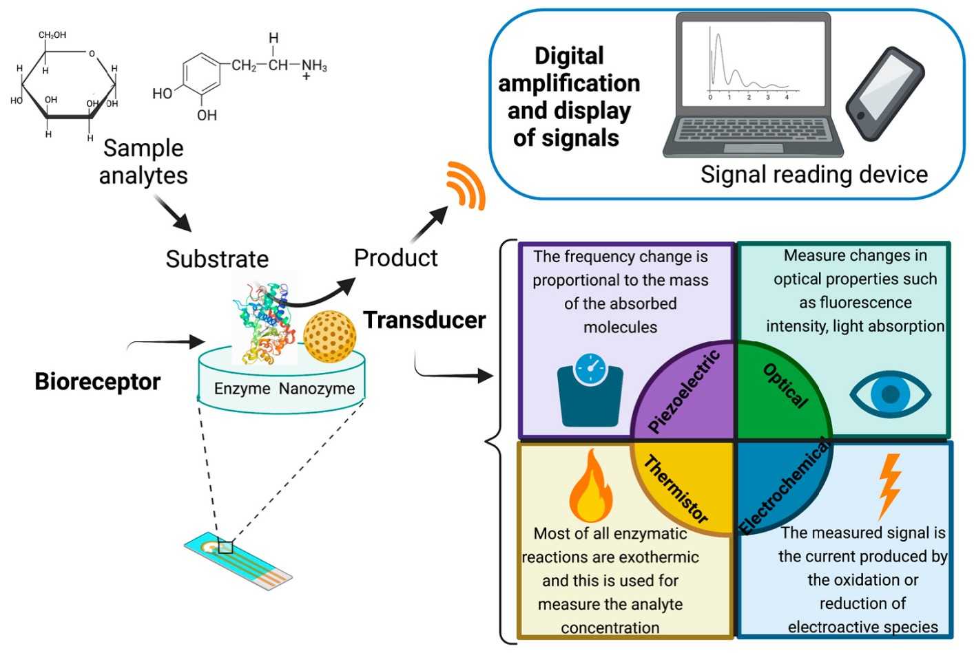
Figure 1. Main biosensing elements and their mechanistic role. (Alvarado-Ramírez et al., 2023)
A biosensor combines a biorecognition element, transducer, signal-processing method, and interpretation rule. Antibodies, aptamers, nucleic-acid probes, or receptors capture the analyte. An enzyme may be the recognition element itself, but in many diagnostic formats it is a reporter attached to a secondary binder. Catalytic turnover converts one binding event into many product molecules, increasing signal.
The CDx context adds a clinical decision boundary. Sensitivity must be accompanied by selectivity, precision, stability, traceable controls, and evidence that the reported category identifies the intended therapy population. A low-cost or portable readout does not relax these requirements. The total system includes sample handling, calibration, software, user steps, and result interpretation.
| Architecture | Enzyme role | Readout | Development concern |
|---|---|---|---|
| Sandwich immunosensor | Reporter on detection antibody | Colorimetric, fluorescent, chemiluminescent, electrochemical | Conjugate ratio and nonspecific binding |
| Competitive sensor | Reporter signal inversely related to analyte | Optical or electrochemical | Dynamic range and intuitive interpretation |
| Enzymatic metabolite sensor | Enzyme directly converts analyte | Current, potential, color, or fluorescence | Interfering substrates and cofactor dependence |
| Nucleic-acid sensor | Enzyme amplifies or cleaves after target recognition | Fluorescence, lateral flow, electrochemical | Off-target amplification and contamination |
| Cascade sensor | Multiple enzymes amplify sequentially | High-gain optical or electrical output | Background amplification and timing control |
Horseradish peroxidase offers fast turnover and diverse substrates, while alkaline phosphatase can provide stable activity under different buffer conditions. The right choice depends on substrate stability, required range, sample matrix, instrument, reaction time, and stopping method.
Maximum turnover is not always desirable. A very fast reporter can saturate early, compress the dynamic range, and magnify timing differences. Enzyme–substrate signal-system optimization balances sensitivity, background, linearity, and read-window robustness.
Attaching an enzyme to an antibody or surface can alter both partners. Random coupling may modify residues near an antigen-binding site, block the enzyme active site, create heterogeneous label ratios, or promote aggregation. Oriented or site-selective strategies can improve consistency but add process complexity. Free enzyme activity and unconjugated antibody affinity should therefore be supplemented by characterization of the final conjugate.
Surface density also matters. Crowding can limit analyte access or substrate diffusion; sparse coverage can reduce signal. Hydrophobic adsorption may denature proteins, while covalent attachment or affinity capture changes orientation and leaching risk. Relevant support includes enzyme–antibody conjugate development, HRP conjugation, and alkaline-phosphatase conjugation.
Nonspecific binding or spontaneous substrate conversion is amplified along with true binding, reducing discrimination near the cutoff.
Diffusion through membranes, microchannels, or crowded surfaces can make apparent kinetics depend on device geometry and sample viscosity.
Signal may continue to develop until stopped or read. User, instrument, and temperature variation can shift the reported result.
Additional risks include endogenous enzyme activity, colored or electroactive interferents, substrate depletion, hook effects, electrode fouling, and lot variation in membranes or conjugates. Controls should challenge the same flow path and reaction environment as the clinical sample.
Whole blood, plasma, serum, tissue lysate, and other specimens impose different constraints. Viscosity can alter flow; hemoglobin can affect optical or electrochemical readout; endogenous peroxidases and phosphatases can create background; heterophilic antibodies can bridge immunoreagents; and medications or metabolites can interfere with electrodes or substrates.
Developers should test individual interferents and representative clinical matrices because pooled or artificial samples may hide patient-to-patient effects. Blocking chemistry, sample pretreatment, reference channels, dilution, or enzyme engineering may be required. Dilution reduces interferents but also reduces the biomarker. Assay interference and matrix-effect evaluation and enzyme inhibitor-tolerance optimization can separate reagent limitations from device effects.
Validation should address accuracy, precision, analytical sensitivity and specificity, reportable range, cutoff behavior, interference, carryover, specimen stability, reagent and sensor lots, reader instruments, operators, and environmental conditions. Point-of-care use may add usability, training, connectivity, and result-transmission risks.
Clinical evidence must use the finalized test or a justified bridged version. If a research biosensor generated trial enrollment data, changes to the binder, enzyme conjugate, substrate, cartridge, reader, algorithm, or cutoff can require bridging. The label must state the intended specimen, therapy, population, and limitations. Because the regulatory landscape evolves, developers should verify current agency requirements and should not describe an emerging biosensor as an authorized CDx without a matching therapeutic claim.
Engineering may improve thermal or pH stability, substrate selectivity, catalytic efficiency, resistance to immobilization, tolerance to matrix components, or expression yield. The screen should use the conjugated or immobilized context whenever possible. A variant optimized in free solution may behave differently after surface attachment.
Tradeoffs require explicit evaluation. Higher activity can narrow the read window; improved stability may reduce turnover; altered surface charge can improve immobilization but increase nonspecific adsorption. Enzyme engineering for CDx and glycerol-free and lyo-ready enzyme development are relevant when device storage or dry-format integration is the limiting factor.
Lateral-flow and membrane-assay enzyme signal support can help evaluate compact formats without assuming that feasibility performance transfers directly to clinical use.
Whatever platform or specimen is selected, development should begin with a written link between intended use and analytical requirements. Define the patient population, biomarker, specimen, treatment decision, reportable result, turnaround expectation, and use environment. Then identify the failure modes that could change classification: target loss, nonspecific signal, amplification bias, reagent drift, interference, software error, or an invalid result that delays therapy. This risk map determines which enzyme attributes and assay controls deserve the most attention.
Feasibility experiments should include representative clinical material as early as possible. Purified templates and synthetic controls are valuable for isolating variables, but they do not reproduce fixation damage, low tumor fraction, endogenous inhibitors, sample heterogeneity, or extraction carryover. A staged study can begin with controlled materials, add individual challenges, and then confirm performance in specimens spanning the intended range. Samples near the cutoff are especially informative because small shifts in recovery, background, or signal can change the treatment category.
Critical enzymes should be specified by more than catalog activity. Identity, purity, concentration, specific activity, contaminating nuclease or protease limits, formulation, storage, and functional performance may all be relevant. The release method should use conditions that predict performance in the diagnostic reaction. When the vendor activity assay and CDx chemistry differ substantially, an assay-level incoming or bridging test can provide a more direct control. Multiple lots should be evaluated before pivotal use so the acceptance range reflects manufacturing variation rather than one favored batch.
Robustness studies intentionally vary parameters that will move in practice: reaction time and temperature, pipetting, sample input, operator, instrument, reagent lot, shipping excursion, and storage duration. Interference studies should use justified concentrations and combinations of endogenous substances, collection additives, medications, and process residuals. Controls must fail when the vulnerable step fails; an abundant control target may remain positive even when a low-copy clinical target is lost.
Finally, document changes across the full measurement system. A new enzyme lot, buffer, primer pool, conjugation process, extraction kit, instrument, or software version can alter analytical performance even if the intended use is unchanged. Risk-based comparability should focus on the attributes most likely to affect the cutoff and claimed range. Preserving retained samples, reference materials, version history, and a predefined bridging strategy makes lifecycle improvements possible without breaking the connection to the clinical evidence.
These questions keep development centered on the treatment decision rather than isolated technical metrics. They also create a common language for biomarker, clinical, regulatory, quality, manufacturing, and supplier teams.
Enzyme-based biosensors can provide sensitive, compact, and rapid biomarker measurement, but catalytic gain also amplifies background, timing, and matrix effects. In CDx development, the enzyme, conjugate, surface, substrate, reader, algorithm, and therapeutic claim must be designed as one system. Careful validation and restrained regulatory language are essential as biosensor applications move from promising prototypes toward treatment-linked diagnostics.
Creative Enzymes supports diagnostic enzyme selection, engineering, formulation, conjugation, analytical evaluation, and scale-up for molecular and immunoassay CDx workflows.
