
Uric acid is the principal end product of purine metabolism in humans. At physiological pH, it is present predominantly as urate, although clinical chemistry assays and laboratory reports commonly use the term “uric acid.” Its concentration reflects the balance among purine production, dietary and cellular sources, renal excretion, and intestinal elimination.
Most enzymatic uric acid methods use uricase, also known as urate oxidase, as the recognition enzyme. The reaction can be monitored through the decrease in urate absorbance or coupled to a hydrogen peroxide reporter system. Method development therefore involves more than selecting uricase alone: enzyme stability, reporter chemistry, sample blanking, interference control, calibration, and analyzer compatibility must be considered together.
Enzymes supplies diverse uricase from multiple microbial and recombinant sources, along with coupling enzymes and chromogenic substrates essential for uric acid assay development, and a complete uric acid assay kit for diagnostic reagent development, metabolic research, method evaluation, and compatible laboratory testing workflows. This integrated product range supports projects from reaction design and raw-material screening to method verification and routine testing.
Humans do not have functional endogenous uricase and therefore do not enzymatically convert urate to allantoin as the final step of purine degradation. Circulating urate is cleared mainly through renal and intestinal pathways.
Uric acid measurement may support laboratory evaluation or research involving:
A uric acid result is not diagnostic by itself. Interpretation depends on the specimen, reference interval, clinical context, medications, renal function, and other laboratory findings.
“Uric acid” and “urate” are often used interchangeably in routine laboratory language, but they describe different protonation states of the same chemical system. At physiological pH, the urate form predominates.
An assay should define whether it reports concentration in serum, plasma, urine, or another validated matrix. Serum and urine applications can require substantially different dilution, calibration, linearity, and sample-blank strategies.
The reported result should also specify:
The assay measures total uric acid or urate recognized under the defined reaction conditions. It does not distinguish the metabolic or clinical cause of an abnormal concentration.
Uricase provides the biochemical specificity in both direct and coupled enzymatic methods. The main difference is whether the assay measures disappearance of urate or formation of hydrogen peroxide.
| Assay Architecture | Measured Change | Principal Components | Development Priorities |
|---|---|---|---|
| Direct uricase ultraviolet method | Decrease in the native ultraviolet absorbance of urate | Uricase, buffer, sample blank, calibrator, and controls | Background absorbance, wavelength accuracy, uricase completeness, blank correction, and spectral interference |
| Uricase-peroxidase colorimetric method | Formation of a colored product through hydrogen peroxide | Uricase, peroxidase, chromogenic substrates, buffer, calibrator, and controls | Reporter compatibility, peroxide recovery, reducing-substance interference, reagent blank, and color stability |
| Uricase-based electrochemical method | Hydrogen peroxide production, oxygen consumption, or another electrode response | Uricase, electrode system, immobilization matrix, mediator or reporter components | Enzyme immobilization, electrode selectivity, signal drift, fouling, oxygen dependence, and operational stability |
Uricase, or urate oxidase, catalyzes the oxygen-dependent oxidation of urate. The immediate enzymatic product is an oxidized urate intermediate, while hydrogen peroxide is generated for use in a coupled detection system.
Analytical reaction: Urate + O2 + H2O → Oxidized urate intermediate + H2O2
The oxidized intermediate can undergo subsequent non-enzymatic or enzyme-assisted conversion toward allantoin. For routine assay design, the relevant analytical events are the loss of urate absorbance and the stoichiometric generation of hydrogen peroxide.
In a coupled colorimetric method, peroxidase uses uricase-generated hydrogen peroxide to oxidize compatible reporter substrates. The resulting dye concentration is measured photometrically and related to uric acid concentration through calibration.
General reporter reaction: H2O2 + reduced reporter substrates → oxidized colored product + H2O
The reporter chemistry determines the final measurement wavelength, color yield, blank behavior, sensitivity, and vulnerability to reducing substances. A general uricase-peroxidase principle does not prescribe one specific chromogen pair or reagent formulation.
Ascorbate can consume hydrogen peroxide or reduce oxidized reporter products, potentially producing a negative bias in peroxide-dependent methods. Ascorbate oxidase may be evaluated as an auxiliary component to reduce this effect before or during signal generation.
Its inclusion is method-dependent. Enzyme concentration, reaction timing, oxygen demand, reagent stability, and interactions with other components must be assessed in the complete formulation. Ascorbate oxidase does not eliminate every source of interference.
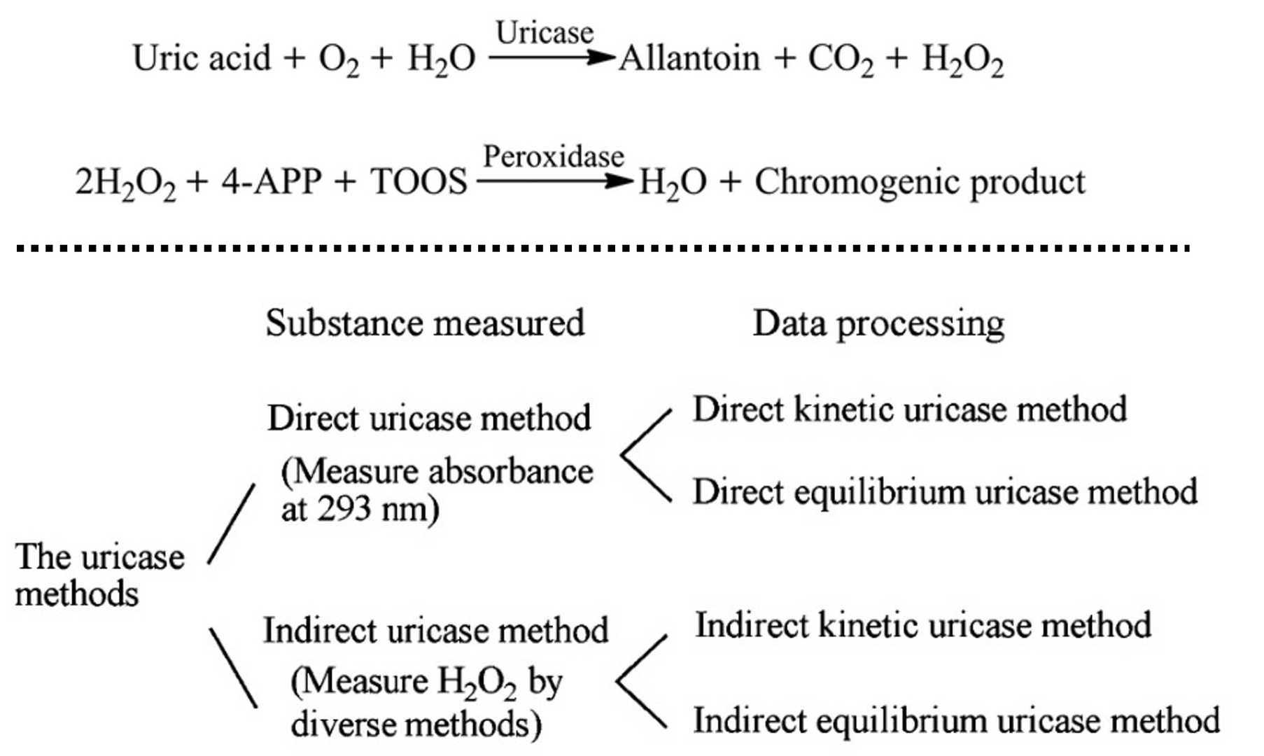 Figure 1. (Top) Mechanism of uric acid assay. (Wen et al., 2019) (Bottom) The classification of the uricase methods for serum uric acid assay. (Zhao et al., 2019)
Uric acid assay systems may combine a recognition enzyme with reporter enzymes, interference-control components, calibrators, and controls. The required components depend on whether the method uses direct ultraviolet measurement, peroxide-dependent color formation, or an electrochemical signal.
| Component | Role in the Assay | Important Selection Criteria |
|---|---|---|
| Uricase / urate oxidase | Recognition enzyme for the selective oxidation of urate | Source, biochemical identity, activity method, specific activity, substrate affinity, pH profile, purity, formulation, and storage stability |
| Peroxidase | Conversion of uricase-generated hydrogen peroxide into a measurable reporter signal | Activity, purity, reporter compatibility, residual interfering activities, formulation compatibility, and stability |
| Ascorbate oxidase | Optional reduction of ascorbate interference in peroxide-dependent methods | Ascorbate-removal capacity, reaction timing, oxygen dependence, pH compatibility, and stability in the complete reagent |
| Uric acid calibrators | Establishment of the relationship between analytical response and uric acid concentration | Assigned value, matrix, traceability, commutability, homogeneity, reconstitution behavior, and stability |
| Quality control materials | Monitoring of calibration status, precision, reagent performance, and analytical drift | Concentration levels, matrix, target values, stability, homogeneity, and compatibility with the measurement procedure |
| Complete uric acid assay kits | Configured reagent systems integrating enzymatic recognition, signal generation, calibration, and application parameters | Assay principle, specimen type, analyzer compatibility, measuring range, calibration model, interference profile, packaging, and storage requirements |
Product selection should be based on the complete measurement procedure. Enzyme activity, reporter chemistry, calibrator matrix, analyzer settings, sample type, and stability requirements should be evaluated as an integrated system.
Uric acid assays can be affected by both enzyme-related and detection-related interference. The relevant interference profile depends strongly on whether the method uses direct ultraviolet absorbance or peroxide-dependent color formation.
| Factor | Possible Effect | Development Response |
|---|---|---|
| Ascorbate and other reducing compounds | Consumption of hydrogen peroxide or reduction of the colored reporter product | Evaluate sample blanking, reaction sequencing, alternative reporter chemistry, or a compatible interference-control enzyme |
| Bilirubin | Spectral interference or chemical interaction with the reporter system | Test conjugated and unconjugated bilirubin across the intended concentration range |
| Hemolysis | Background absorbance, released cellular substances, or peroxidase-like effects | Establish method-specific hemolysis limits rather than relying on another reagent’s claims |
| Lipemia and turbidity | Light scattering and elevated sample background | Evaluate bichromatic measurement, sample blanking, or validated clearing strategies |
| Endogenous catalase or catalase contamination | Loss of hydrogen peroxide and negative bias in coupled methods | Control enzyme purity and investigate peroxide recovery in the final formulation |
| Peroxidase-active or redox-active substances | Non-specific color formation or reporter suppression | Perform structured interference studies using the complete reagent system |
| Specimen-type differences | Matrix-dependent recovery, dilution, or bias | Validate serum, plasma, and urine separately and define acceptable anticoagulants |
| High urate concentration | Nonlinearity, substrate depletion, incomplete conversion, or solubility-related handling challenges | Establish the analytical measuring range and a validated sample-dilution procedure |
Calibration materials should be selected according to the intended matrix and measurement procedure. An aqueous urate solution may support early reaction development, but it is not automatically equivalent to a matrix-based clinical calibrator.
Calibration development may need to address:
Before a reagent or kit is assigned performance claims, analytical evaluation should include:
Project scope, technical specifications, testing requirements, deliverables, and supply format can be defined according to the intended assay and development stage.
Discuss a Uric Acid Assay Requirement
Q1. Are uric acid and urate the same analyte?
Q2. What is the principal enzyme used in a uric acid assay?
Q3. What is the difference between direct and coupled uricase methods?
Q4. Why can ascorbate interfere with a uricase-peroxidase assay?
Q5. Can the same reagent be used for serum, plasma, and urine?
Q6. What components may be required for a uric acid assay?
Q7. What should be considered when selecting uricase?
Q8. Does an elevated uric acid result diagnose gout?
Q9. Can a new reagent reproduce an existing commercial assay formulation?
