
Tissue and liquid biopsy can reveal treatment-relevant tumor biology through different sampling windows. Tissue provides morphology, spatial context, and direct tumor material. Plasma cell-free DNA offers a less invasive sample that may capture DNA shed from multiple lesions and permit repeat collection. Their limitations—and their enzyme requirements—are not the same.
This guide compares tissue and liquid-biopsy CDx workflows from preanalytics through extraction, amplification, library preparation, error control, and interpretation. It emphasizes why polymerase, reverse-transcriptase, ligase, and processing-enzyme selection should follow specimen physics and the intended therapy claim.
A tissue section can confirm tumor presence, estimate tumor content, preserve architecture, and support protein or nucleic-acid testing. Yet one biopsy samples one location and time, may contain necrosis or low tumor cellularity, and can be damaged by fixation. Repeat invasive collection may be difficult.
Liquid biopsy most commonly analyzes circulating tumor DNA within total plasma cell-free DNA. It can sample material from multiple disease sites, but tumor-derived molecules may be rare or absent because shedding depends on burden, location, biology, treatment, and collection timing. Plasma is therefore not simply “tissue without a biopsy.” The intended-use label and evidence determine how either result should guide treatment.
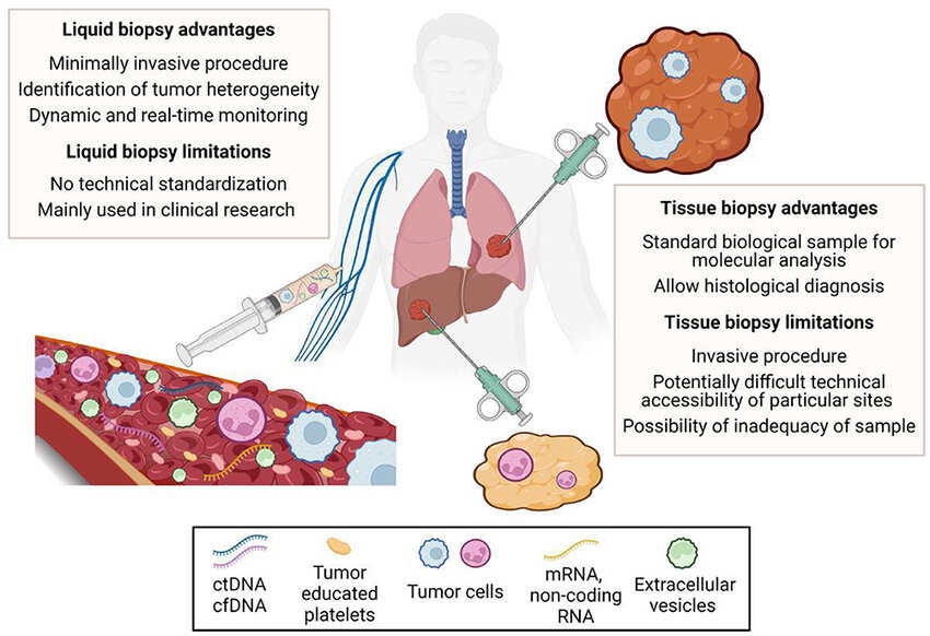
Figure 1. Comparison between tissue and liquid biopsy: advantages and limitations. (Nigro et al., 2023)
| Dimension | Tissue biopsy | Plasma liquid biopsy |
|---|---|---|
| Biological context | Morphology and local tumor microenvironment | Systemic mixture of cell-free DNA from multiple sources |
| Tumor analyte fraction | Often higher after tumor enrichment | Can be very low and variable |
| Preanalytics | Ischemia, fixation, sectioning, decalcification, tumor content | Tube type, processing delay, centrifugation, hemolysis, plasma storage |
| Nucleic-acid condition | FFPE damage, crosslinks, fragmentation | Short fragments, low mass, background wild-type DNA |
| Sampling limitation | Spatial heterogeneity and finite tissue | Variable shedding and dilution in total cfDNA |
| Interpretive confounder | Normal-cell admixture and fixation artifacts | Clonal hematopoiesis and low-molecule sampling |
| Repeat access | May require another procedure | Serial blood draws are generally more feasible |
| Additional modalities | IHC, ISH, morphology, DNA and RNA | Primarily molecular; circulating cells/proteins are separate workflows |
Formalin can fragment and crosslink nucleic acids and create sequence artifacts. Decalcification can further damage material. Plasma delays can release genomic DNA from blood cells, diluting tumor signal, while incorrect centrifugation or freeze–thaw handling changes yield.
Enzyme engineering cannot fully recover destroyed targets. Collection and extraction controls should be optimized first, then enzymes selected for the remaining fragment lengths, inhibitors, and input range.
FFPE-derived DNA often consists of short, chemically modified fragments. Amplicons should be appropriately sized, and polymerases should tolerate relevant extraction carryover while maintaining specificity. High processivity cannot compensate for templates broken between primer sites. Damage-associated substitutions may require enzymatic repair strategies, molecular controls, or bioinformatic filters, each validated for possible bias.
RNA from tissue can support fusion or expression testing but is especially vulnerable to degradation. Reverse-transcription temperature, priming, RNase control, and short target design influence recovery. AdvSTART Reverse Transcriptase and one-step RT-qPCR development are relevant for feasibility, followed by testing across representative RNA integrity and fixation conditions.
Cell-free DNA is short and low in mass, and tumor alleles may be greatly outnumbered by wild-type molecules. Extraction recovery, adapter ligation, and early amplification efficiency can determine how many original molecules remain observable. High activity alone is not enough; low bias and a controlled error profile are vital.
NGS workflows may use end repair, A-tailing, ligation, and library amplification. Unique molecular identifiers are most effective when attached before extensive copying, but inefficient ligation loses molecules before they can be counted. T4 DNA Ligase (Rapid) and NGS library-preparation enzyme-system development can support low-input studies. PCR workflows may prioritize hot-start specificity and short targets; Taq HS DNA Polymerase is a representative candidate.
Fixation damage and deamination can resemble variants. Controls, replicate evidence, repair, and filters must be evaluated without erasing true calls.
Low tumor fraction makes polymerase and sequencing errors more visible; wild-type DNA and clonal hematopoiesis complicate attribution.
UMIs and duplex evidence can distinguish original molecules from copies, but cannot recover molecules lost during collection or library preparation.
Uracil-processing enzymes may reduce certain deamination artifacts or carryover in compatible workflows, but they can also alter templates or chemistry. Every error-control intervention needs assay-level verification across representative variant classes and allele fractions.
Analytical limit of detection should be expressed in terms that reflect the workflow: allele fraction, input mass, and preferably target-molecule availability. At very low input, replicate results vary because of sampling even when chemistry is perfect. Plasma sensitivity also changes with biological shedding. A negative plasma result can mean no target alteration, insufficient tumor DNA, or failure to sample the relevant disease signal.
Whether tissue follow-up is recommended depends on the specific test label, disease, therapy, specimen adequacy, and clinical setting. It should not be stated as a universal rule. Tissue negatives also have limitations, including spatial sampling and insufficient tumor. Reports and clinical protocols should explain invalid, insufficient, below-limit, and not-detected categories clearly.
A plasma claim cannot be established solely with contrived DNA in buffer, and a tissue claim cannot rely only on high-quality frozen DNA. Studies should cover collection devices, processing times, storage, extraction lots, input range, endogenous interferents, tumor fraction, variant types, and near-cutoff concentrations. Contrived materials are useful but should complement representative clinical specimens.
Comparing tissue and plasma requires careful timing and biological interpretation. Discordance can reflect heterogeneity, therapy effects, shedding, clonal hematopoiesis, or analytical error. Agreement statistics should be accompanied by analysis of these causes. Interference and matrix evaluation and nucleic-acid extraction enzyme-system optimization can support specimen-specific controls.
Tissue and plasma workflows may share enzymes but use different buffer, concentration, and workflow constraints. Low-input plasma libraries are sensitive to adsorption and dead volume; tissue extracts may carry salts, ethanol, paraffin residues, or inhibitors. Premixes simplify handling but must remain stable with all cofactors and oligonucleotides.
Real-time and justified accelerated stability studies should use final containers and test assay-level performance. Shipping excursions, freeze–thaw cycles, on-board stability, and reconstitution matter. Diagnostic enzyme stability and shelf-life testing and lyo-ready enzyme development are relevant when decentralized or cartridge formats are planned.
The CDx technology platform can integrate specimen and method selection under one intended-use strategy.
Whatever platform or specimen is selected, development should begin with a written link between intended use and analytical requirements. Define the patient population, biomarker, specimen, treatment decision, reportable result, turnaround expectation, and use environment. Then identify the failure modes that could change classification: target loss, nonspecific signal, amplification bias, reagent drift, interference, software error, or an invalid result that delays therapy. This risk map determines which enzyme attributes and assay controls deserve the most attention.
Feasibility experiments should include representative clinical material as early as possible. Purified templates and synthetic controls are valuable for isolating variables, but they do not reproduce fixation damage, low tumor fraction, endogenous inhibitors, sample heterogeneity, or extraction carryover. A staged study can begin with controlled materials, add individual challenges, and then confirm performance in specimens spanning the intended range. Samples near the cutoff are especially informative because small shifts in recovery, background, or signal can change the treatment category.
Critical enzymes should be specified by more than catalog activity. Identity, purity, concentration, specific activity, contaminating nuclease or protease limits, formulation, storage, and functional performance may all be relevant. The release method should use conditions that predict performance in the diagnostic reaction. When the vendor activity assay and CDx chemistry differ substantially, an assay-level incoming or bridging test can provide a more direct control. Multiple lots should be evaluated before pivotal use so the acceptance range reflects manufacturing variation rather than one favored batch.
Robustness studies intentionally vary parameters that will move in practice: reaction time and temperature, pipetting, sample input, operator, instrument, reagent lot, shipping excursion, and storage duration. Interference studies should use justified concentrations and combinations of endogenous substances, collection additives, medications, and process residuals. Controls must fail when the vulnerable step fails; an abundant control target may remain positive even when a low-copy clinical target is lost.
Finally, document changes across the full measurement system. A new enzyme lot, buffer, primer pool, conjugation process, extraction kit, instrument, or software version can alter analytical performance even if the intended use is unchanged. Risk-based comparability should focus on the attributes most likely to affect the cutoff and claimed range. Preserving retained samples, reference materials, version history, and a predefined bridging strategy makes lifecycle improvements possible without breaking the connection to the clinical evidence.
These questions keep development centered on the treatment decision rather than isolated technical metrics. They also create a common language for biomarker, clinical, regulatory, quality, manufacturing, and supplier teams.
Tissue and liquid biopsy are complementary rather than interchangeable CDx specimens. Tissue offers tumor context and often more direct analyte access; plasma offers repeatable, minimally invasive sampling but may contain very few tumor molecules. Enzyme systems should be designed around these realities, with specimen-specific validation, controlled error mitigation, and interpretation rules aligned to the exact therapy claim.
Creative Enzymes supports diagnostic enzyme selection, engineering, formulation, conjugation, analytical evaluation, and scale-up for molecular and immunoassay CDx workflows.
