
Molecular diagnostics and immunoassays answer different questions about tumor biology and treatment response. Molecular methods measure DNA or RNA variants, copy-number changes, fusions, expression, or composite genomic signatures. Immunoassays generally measure proteins, their abundance, localization, or modification in cells and tissues. Either approach can support a companion diagnostic when its result is essential to the safe and effective use of a therapy.
The platform choice should follow the therapeutic mechanism, biomarker biology, specimen pathway, and intended-use claim. This guide compares the two families across analyte access, enzyme roles, analytical risks, workflow, interpretation, and co-development strategy.
DNA is relatively stable and can reveal inherited or acquired variants, but a genomic alteration does not always establish that a protein is expressed or functionally active. RNA can capture fusions and transcriptional activity, although it is more labile. Protein measurement is closer to phenotype and can preserve spatial context in tissue, yet antibody binding, fixation, heterogeneity, and scoring introduce distinct variables.
A therapy directed at a specific activating mutation may align naturally with PCR or sequencing. A therapy whose response depends on surface-protein abundance or immune-marker localization may require immunohistochemistry. Some biomarkers can be measured at more than one layer, but the results are not automatically interchangeable. Concordance studies must address the biological and technical reasons for disagreement.
| Dimension | Molecular diagnostics | Immunoassays |
|---|---|---|
| Primary analyte | DNA or RNA | Protein or protein-associated structure |
| Representative formats | PCR, qPCR, digital PCR, NGS, in situ hybridization | IHC, ELISA, chemiluminescent assays, lateral flow, biosensors |
| Typical output | Variant, fusion, copy number, expression, genomic score | Intensity, concentration, proportion, localization, categorical score |
| Enzyme roles | Extraction, reverse transcription, amplification, ligation, library preparation | Reporter generation, conjugate systems, substrate conversion, sample processing |
| Key preanalytics | Collection tube, ischemia, extraction, nucleic-acid quality | Fixation, antigen retrieval, tissue processing, epitope preservation |
| Major interpretation risks | Low allele fraction, sequencing artifacts, bioinformatics, contamination | Antibody specificity, staining heterogeneity, observer or image-analysis variability |
| Multiplexing | High with targeted panels or NGS | Usually lower, though multiplex imaging and arrays expand capacity |
| Information retained | Sequence-level detail; spatial context often lost | Protein and tissue context; sequence detail not provided |
Hot-start polymerases suppress extension during setup and early heating, reducing primer-dimers and nonspecific products. This is valuable in multiplex CDx assays where many primers compete. Other relevant attributes include extension rate, processivity, GC tolerance, fidelity, probe compatibility, and resistance to extraction carryover.
Enzyme concentration should be optimized with magnesium, primers, probes, dNTPs, and cycle conditions. Increasing polymerase can recover weak signal but can also raise background or alter multiplex balance. Taq HS DNA Polymerase is a representative starting material; suitability must be verified in the final assay.
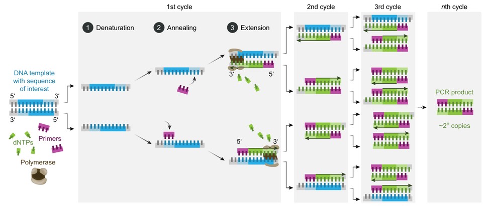
Molecular workflows can use proteases or nucleases during preparation, reverse transcriptase for RNA targets, DNA polymerases for amplification, and ligases or end-repair enzymes for NGS libraries. Each transformation can introduce loss, bias, error, or contamination. A qPCR result depends on primer–probe specificity, polymerase behavior, amplification efficiency, threshold rules, and controls. An NGS result additionally depends on library complexity, coverage, sequence-quality filters, and bioinformatics.
Relevant development paths include PCR and qPCR enzyme/premix development, one-step RT-qPCR master mix development, and NGS library-preparation enzyme systems. Representative raw materials include Taq HS DNA Polymerase, AdvSTART Reverse Transcriptase, and T4 DNA Ligase (Rapid).
Horseradish peroxidase and alkaline phosphatase are common reporter enzymes. After an antibody recognizes the biomarker, the reporter converts substrate into a colored, fluorescent, chemiluminescent, or electrochemical signal. Catalytic amplification improves sensitivity but also magnifies nonspecific binding and timing variation. Conjugation ratio, antibody affinity, enzyme activity, substrate kinetics, washes, incubation, temperature, and readout all shape the result.
HRP conjugation, alkaline-phosphatase conjugation, and enzyme–substrate signal optimization can be evaluated when a reporter system limits sensitivity, background, or precision. The final conjugate should be characterized as a material in its own right because free enzyme activity does not predict antibody binding or surface behavior.
Establish extraction recovery, analytical sensitivity, inclusivity, exclusivity, allele-fraction behavior, contamination control, and algorithm performance.
Establish antibody specificity, staining or signal range, precision, interference, hook effects, scoring reproducibility, and calibrator traceability.
Evaluate accuracy, precision, cutoff behavior, invalid rates, specimen stability, reagent lots, instruments, operators, and robustness around intended use.
Method comparison alone is insufficient when no perfect reference exists. Discordant analysis should use orthogonal evidence and account for tumor heterogeneity, analyte decay, sampling, and each method’s detection limit. Clinical validation must show that the finalized test identifies the population described in the therapeutic labeling.
Molecular cutoffs may involve variant allele frequency, copy-number thresholds, expression scores, or multivariate algorithms. Immunoassay cutoffs may use staining intensity, percentage of positive cells, combined scores, or concentration. In both cases, a cutoff converts continuous and uncertain biology into a treatment category. Its selection must be prespecified and supported with analytical and clinical evidence.
Borderline samples are critical. Small changes in tumor content, extraction yield, amplification efficiency, staining intensity, or reader interpretation can cross the decision boundary. Precision profiles around the cutoff, not only at high and low controls, reveal this risk. If image analysis or bioinformatics is part of the device, version control, cybersecurity, data integrity, and change assessment become part of lifecycle management.
Combined testing can resolve different parts of a biological question—for example, genomic alteration plus protein expression, or tumor genotype plus an immune-context marker. It can also consume more tissue, extend turnaround time, and complicate interpretation. A sequential strategy may preserve material: a rapid targeted test first, followed by broader profiling or protein assessment when needed.
If results are combined into one CDx decision, the algorithm, missing-data rules, order of testing, and conflict resolution must be validated. Two independently useful tests do not automatically create a validated composite. Developers should also determine whether both are essential to the therapy claim or whether one is exploratory, prognostic, or complementary.
The companion diagnostic technology platform provides a framework for evaluating molecular profiling and protein-expression routes under a common intended-use strategy.
Whatever platform or specimen is selected, development should begin with a written link between intended use and analytical requirements. Define the patient population, biomarker, specimen, treatment decision, reportable result, turnaround expectation, and use environment. Then identify the failure modes that could change classification: target loss, nonspecific signal, amplification bias, reagent drift, interference, software error, or an invalid result that delays therapy. This risk map determines which enzyme attributes and assay controls deserve the most attention.
Feasibility experiments should include representative clinical material as early as possible. Purified templates and synthetic controls are valuable for isolating variables, but they do not reproduce fixation damage, low tumor fraction, endogenous inhibitors, sample heterogeneity, or extraction carryover. A staged study can begin with controlled materials, add individual challenges, and then confirm performance in specimens spanning the intended range. Samples near the cutoff are especially informative because small shifts in recovery, background, or signal can change the treatment category.
Critical enzymes should be specified by more than catalog activity. Identity, purity, concentration, specific activity, contaminating nuclease or protease limits, formulation, storage, and functional performance may all be relevant. The release method should use conditions that predict performance in the diagnostic reaction. When the vendor activity assay and CDx chemistry differ substantially, an assay-level incoming or bridging test can provide a more direct control. Multiple lots should be evaluated before pivotal use so the acceptance range reflects manufacturing variation rather than one favored batch.
Robustness studies intentionally vary parameters that will move in practice: reaction time and temperature, pipetting, sample input, operator, instrument, reagent lot, shipping excursion, and storage duration. Interference studies should use justified concentrations and combinations of endogenous substances, collection additives, medications, and process residuals. Controls must fail when the vulnerable step fails; an abundant control target may remain positive even when a low-copy clinical target is lost.
Finally, document changes across the full measurement system. A new enzyme lot, buffer, primer pool, conjugation process, extraction kit, instrument, or software version can alter analytical performance even if the intended use is unchanged. Risk-based comparability should focus on the attributes most likely to affect the cutoff and claimed range. Preserving retained samples, reference materials, version history, and a predefined bridging strategy makes lifecycle improvements possible without breaking the connection to the clinical evidence.
These questions keep development centered on the treatment decision rather than isolated technical metrics. They also create a common language for biomarker, clinical, regulatory, quality, manufacturing, and supplier teams.
Molecular diagnostics and immunoassays are not competing versions of the same measurement. They interrogate different biological layers and carry different specimen, reagent, workflow, and interpretation risks. A defensible CDx platform is the one that measures the therapy-relevant biomarker in the intended population with controlled uncertainty and a feasible clinical workflow.
Creative Enzymes supports diagnostic enzyme selection, engineering, formulation, conjugation, analytical evaluation, and scale-up for molecular and immunoassay CDx workflows.
