Background
While HRP and AP dominate conventional immunoassay detection, β-galactosidase (β-Gal), urease, and oxidases (glucose oxidase, lactate oxidase, cholesterol oxidase) occupy specialized niches where their unique catalytic mechanisms deliver distinct analytical advantages. β-Gal offers femtomole-level sensitivity through fluorogenic substrate turnover; urease drives pH-shift and electrochemical biosensors without requiring optical readouts; oxidases generate hydrogen peroxide for coupled amplification cascades and amperometric sensor architectures. Creative Enzymes Diagnostic provides dedicated labeling services for all three enzyme classes, tailoring conjugation chemistry and assay optimization to each enzyme's structural requirements and the client's target application.
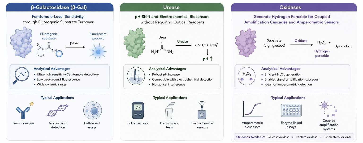 Figure 1. Reporter enzyme selection for specialized diagnostic detection platforms.
Reporter Enzyme Selection
Choosing the appropriate reporter enzyme depends on assay format, detection instrumentation, sensitivity requirements, and sample matrix characteristics. The table below summarizes the key properties of β-galactosidase, urease, and oxidases to guide selection.
| Enzyme |
Source / Structure |
Signal Output |
Key Strengths |
Primary Platforms |
| β-Galactosidase |
E. coli or recombinant; ~465 kDa homotetramer |
Colorimetric (yellow/blue precipitate), fluorescent, chemiluminescent |
Highest sensitivity among common reporters; broad substrate palette; minimal endogenous background in mammalian samples |
Ultra-sensitive ELISA, Western blot, molecular diagnostics, single-molecule detection |
| Urease |
Jack bean or bacterial; ~480 kDa hexamer with Ni2+ active site |
pH shift (phenol red), conductivity change, ammonia detection (ISE), amperometric |
Signal amplification through pH cascade; no optical interference; ideal for electrochemical and colorimetric paper-based assays |
Biosensors, POC dipstick assays, electrochemical immunoassays, urea diagnostic kits |
| Oxidases (GOx, LOx, ChOx) |
Fungal or bacterial; ~160 kDa dimer (GOx), monomeric variants |
H2O2 generation for coupled HRP/AP/ferrocene detection; O2 depletion (Clark electrode) |
Natural specificity for clinically relevant analytes; dual-use as reporter and recognition element; compatible with amperometric electrodes |
Glucose/lactate/cholesterol biosensors, coupled amplification ELISA, wearable sensors |
β-Galactosidase
- High sensitivity: β-Gal's high catalytic turnover with fluorogenic substrates (4-methylumbelliferyl-β-D-galactopyranoside, MUG; resorufin-β-D-galactopyranoside) enables detection limits in the femtomole range—10- to 100-fold lower than standard HRP-TMB systems—making it the preferred reporter for ultra-sensitive cytokine, autoantibody, and nucleic acid hybridization assays
- Colorimetric: Ortho-nitrophenyl-β-D-galactopyranoside (ONPG) yields a soluble yellow product (A420) for quantitative kinetic assays; 5-bromo-4-chloro-3-indolyl-β-D-galactopyranoside (X-Gal) produces an insoluble blue precipitate for membrane-based and histochemical localization studies
- Fluorescent: Fluorescein di-β-D-galactopyranoside (FDG) and similar substrates generate bright fluorescent products with high quantum yield, enabling multiplexed fluorescence detection alongside GFP, Cy-dye, or Alexa Fluor channels without spectral overlap
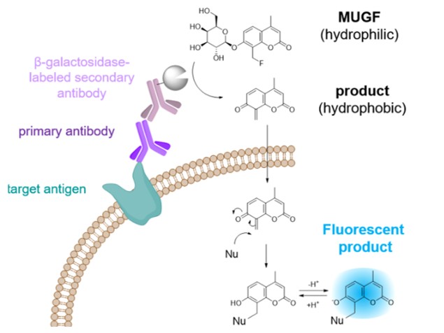 Figure 2. β-galactosidase-catalyzed fluorescent reporter labeling of living cells for sensitive detection of cell surface antigens. (Noguchi et al., 2020)
Urease
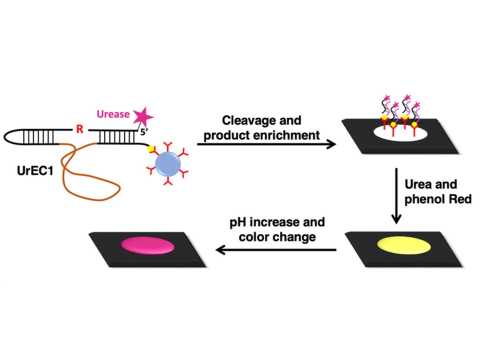 Figure 3. Schematic illustration of the test principle for a colorimetric assay based on EC1 labelled with urease. (Samani et al., 2020)
- Biosensors: Urease catalyzes the hydrolysis of urea to ammonia and carbon dioxide, causing a localized pH increase that can be detected by pH-sensitive dyes (phenol red, bromocresol purple) in paper-based or microfluidic lateral flow devices. This colorimetric pH shift requires no optical excitation source, reducing instrument complexity for point-of-care diagnostics
- Electrochemical assays: The ammonium ions generated by urease activity are quantifiable by ion-selective electrodes (ISE) or amperometric detection of pH change at microfabricated iridium oxide or palladium electrodes. Urease-labeled immunoconjugates enable label-free electrochemical immunoassays with detection limits comparable to optical ELISA
Oxidases
- Glucose oxidase (GOx): Widely deployed as both a recognition element and reporter in glucose biosensors. In immunoassay applications, GOx generates H2O2 that drives a secondary HRP-mediated signal amplification cascade, effectively creating a two-enzyme amplification loop that boosts sensitivity beyond single-enzyme detection
- Lactate oxidase (LOx) and cholesterol oxidase (ChOx): LOx and ChOx provide substrate-specific signal generation for metabolite panels. When conjugated to antibodies or streptavidin, these enzymes serve as reporters in competitive immunoassays for small-molecule drugs and hormones, leveraging the natural specificity of the oxidase for its cognate substrate to minimize cross-reactivity
- Diagnostic biosensors: Oxidase conjugates immobilized on electrode surfaces (carbon nanotube, gold nanoparticle, or graphene-modified electrodes) enable direct amperometric detection of H2O2 or O2 depletion, supporting continuous monitoring, wearable patch sensors, and microfluidic integrated diagnostic platforms
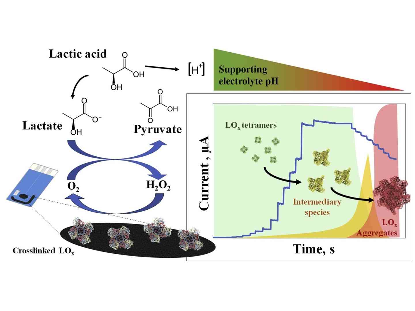 Figure 4. Inhibited enzymatic reaction of crosslinked lactate oxidase through a pH-dependent mechanism. (Cunha-Silva et al., 2019)
Conjugation Strategies
Our labeling services are organized by target substrate rather than by enzyme, ensuring that the conjugation chemistry is optimized for the intended carrier (antibody, protein, or nanoparticle) while preserving the unique cofactor and structural requirements of β-Gal, urease, or oxidase.
Antibody Labeling
- Controlled random coupling via amine-reactive NHS-esters or thiol-maleimide chemistry, with strict optimization of enzyme-to-antibody ratios to prevent aggregation—particularly critical for the large tetrameric β-Gal and hexameric urease, where excessive cross-linking causes precipitate formation and loss of both enzymatic and binding activities
- Site-directed conjugation to antibody Fc glycans (periodate oxidation/reductive amination) or engineered hinge cysteines, directing the bulky enzyme away from the antigen-binding paratope and preserving affinity
- Validation of conjugate monodispersity by size-exclusion chromatography (SEC) and dynamic light scattering (DLS); only fractions with polydispersity index (PDI) 85% activity retention are qualified for release
Protein Labeling
- Streptavidin- and NeutrAvidin-enzyme conjugates for universal biotinylated antibody detection across multiple assay formats; biotin-binding capacity verified by HABA displacement assay to ensure ≥12–14 biotin binding sites per streptavidin tetramer remain functional after conjugation
- Protein A/G enzyme conjugates for species-flexible secondary detection in ELISA, Western blot, and immunoprecipitation workflows; binding to human, mouse, rabbit, and goat IgG subclasses validated by SPR to confirm Fc-binding affinity is preserved
- Custom protein-enzyme fusions (e.g., scFv-β-Gal, nanobody-urease) generated by recombinant expression in E. coli or mammalian systems, with optional linker engineering (flexible Gly-Ser repeats or rigid α-helical spacers) to optimize inter-domain flexibility and reduce steric clash
Nanoparticle Labeling
- Gold nanoparticle (AuNP) conjugation via electrostatic adsorption or covalent thiol linkage, creating enzyme-nanoparticle hybrids with enhanced local enzyme density and amplified signal output; AuNP size (5–40 nm) optimized to balance colloidal stability against sedimentation in microfluidic channels
- Magnetic bead (Fe3O4@SiO2) functionalization for automated immunoassay platforms; magnetic separation enables rapid wash steps and reduced background, with enzyme loading quantified by BCA and activity assays to ensure consistent bead-to-bead performance
- Quantum dot and upconversion nanoparticle coupling for multimodal detection platforms combining enzymatic signal amplification with fluorescence or luminescence barcoding; surface PEGylation evaluated to prevent non-specific binding and nanoparticle aggregation in serum matrices
Assay Optimization
Each reporter enzyme class presents distinct optimization challenges. Our assay development team addresses signal stability, background suppression, and storage formulation to ensure robust diagnostic performance.
Signal Stability
- β-Gal signal stability optimized by substrate selection: MUG and chemiluminescent substrates (e.g., Galacton-Star) provide extended glow kinetics compared to flash-type systems; reaction kinetics monitored in real time to determine the linear accumulation window for endpoint or kinetic readout
- Urease pH-shift assays buffered with weakly buffered or unbuffered indicator systems to maximize pH change magnitude; reaction quenching with acidic or basic stop solutions evaluated to fix endpoint color for batch reading
- Oxidase coupled systems optimized for H2O2 steady-state concentration to prevent HRP suicide inactivation; oxygen availability in sealed microplate wells assessed and compensated by increased headspace or permeable film seals
Background Reduction
- β-Gal background minimized by sample matrix treatment with chloroquine or competitive inhibitors during non-specific binding steps, and by using heat-inactivated or lacZ-deficient assay diluents to suppress any endogenous bacterial enzyme activity in environmental or agricultural samples
- Urease background controlled by rigorous removal of free urea from buffers and by inclusion of urease inhibitors (acetohydroxamic acid) in negative-control wells to establish baseline drift rates
- Oxidase background managed by catalase co-addition to scavenge ambient H2O2 in buffers and sample matrices, and by selection of low-autofluorescence microplates for coupled fluorescence readouts
Storage Stability
- β-Gal conjugates stabilized in buffers containing Mg2+ and K+ (essential cofactors), BSA or trehalose as cryoprotectants, and glycerol (30–50%) for −20°C storage; tetramer dissociation monitored by native PAGE to ensure subunit integrity over 12-month storage
- Urease conjugates protected by Ni2+ and Mn2+ supplementation in formulation buffers; lyophilization in sucrose/trehalose matrices validated for ambient-temperature stability in POC shipping scenarios
- Oxidase conjugates formulated at pH 5.5–7.0 to minimize FAD dissociation from the apoenzyme; amber vials and oxygen-depleted headspace nitrogen purging evaluated for long-term FAD retention in glucose oxidase conjugates
FAQs
Q1. Which reporter enzyme should I choose for maximum sensitivity in ELISA?
A1. β-Galactosidase with a fluorogenic substrate (MUG or Galacton-Star) typically delivers the highest sensitivity, with detection limits 10- to 100-fold lower than HRP-TMB systems. For single-molecule or digital ELISA applications, β-Gal is the established reporter of choice. If your laboratory lacks fluorescence capability, urease with a pH-indicator endpoint can achieve sub-picomolar sensitivity in optimized buffer systems.
Q2. Can urease conjugates be used in standard microplate ELISA readers?
A2. Yes. Urease conjugates are fully compatible with standard absorbance microplate readers. The most common readout uses phenol red (A560) or bromocresol purple as a pH indicator, producing a color change proportional to urease activity. No specialized electrochemical equipment is required for colorimetric urease ELISA. Electrochemical readouts are available as an advanced option for biosensor development.
Q3. Are oxidase conjugates suitable for applications requiring HRP-free detection?
A3. Absolutely. While oxidases are frequently coupled with HRP for amplified signal generation, they can also serve as standalone reporters. Direct amperometric detection of H2O2 at platinum or carbon electrodes, oxygen depletion measured by Clark electrodes, or colorimetric detection via ferrocyanide/ferricyanide redox cycling all enable HRP-free assay architectures. We can design and validate oxidase-only detection schemes based on your instrument platform.
Q4. Do large enzymes like β-Gal and urease cause steric hindrance in sandwich assays?
A4. The large size of β-Gal (~465 kDa) and urease (~480 kDa) can cause steric issues if conjugation is uncontrolled. We mitigate this through (1) site-specific Fc-directed labeling that positions the enzyme away from the antigen-binding site, (2) strict control of degree of labeling (DOL ≤ 2), and (3) use of flexible linker sequences in recombinant fusions. In practice, our optimized β-Gal and urease antibody conjugates perform robustly in standard sandwich ELISA with no detectable hook effect or affinity loss.
Q5. Can you label nanoparticles with more than one enzyme type for multiplexed detection?
A5. Yes. We engineer multi-enzyme nanoparticle conjugates for multiplexed assays. For example, β-Gal and urease can be co-immobilized on magnetic beads, with each enzyme providing a distinct signal channel (fluorescence vs. pH color). Alternatively, different oxidases (GOx, LOx, ChOx) can be patterned on electrode arrays for simultaneous metabolite panel detection. We optimize surface chemistry to prevent enzyme–enzyme interference and ensure uniform co-loading.
Q6. What is the typical timeline for a custom labeling project?
A6. Standard custom conjugation projects require 6–8 weeks: 1–2 weeks for feasibility and chemistry selection, 2–3 weeks for conjugation and purification, and 2–3 weeks for analytical characterization (activity, binding, DOL, purity) and assay validation. Recombinant fusion protein projects extend to 10–12 weeks to accommodate expression and purification. Expedited timelines are available for diagnostic development programs with critical milestones.
Creative Enzymes Diagnostic provides comprehensive labeling services for β-galactosidase, urease, and oxidase conjugates, bridging the gap between specialized enzyme biochemistry and practical diagnostic assay development. Whether your application demands the extreme sensitivity of fluorogenic β-Gal detection, the instrument simplicity of urease pH-shift assays, or the biosensor integration of oxidase electrochemistry, our team delivers optimized conjugates with validated performance and batch consistency.
Contact our business development team today to discuss your reporter enzyme labeling requirements!
References
- Noguchi K, Shimomura T, Ohuchi Y, et al. Β-galactosidase-catalyzed fluorescent reporter labeling of living cells for sensitive detection of cell surface antigens. Bioconjugate Chem. 2020;31(7):1740-1744. doi:10.1021/acs.bioconjchem.0c00180
- Samani SE, Chang D, McConnell EM, Rothenbroker M, Filipe CDM, Li Y. Highly sensitive RNA‐cleaving dnazyme sensors from surface‐to‐surface product enrichment. ChemBioChem. 2020;21(5):632-637. doi:10.1002/cbic.201900575
- Cunha-Silva H, Pires F, Dias-Cabral AC, Arcos-Martinez MJ. Inhibited enzymatic reaction of crosslinked lactate oxidase through a pH-dependent mechanism. Colloids and Surfaces B: Biointerfaces. 2019;184:110490. doi:10.1016/j.colsurfb.2019.110490
Related Services

