Background
The enzyme-substrate reaction is the signal-generating engine of every diagnostic immunoassay. While much attention focuses on antibody specificity and conjugate chemistry, the ultimate sensitivity, dynamic range, and reproducibility of an assay are determined by how efficiently the reporter enzyme converts substrate into detectable product—and how effectively that signal is captured by the readout instrument. Suboptimal substrate selection, incompatible buffer matrices, or mismatched detection modalities can degrade even the finest antibody-enzyme conjugate into a mediocre reagent. Creative Enzymes Diagnostic offers a dedicated Enzyme-Substrate Signal System Optimization service that engineers the entire signal generation cascade, from enzyme and substrate pairing through buffer formulation and detection system integration, to unlock the full analytical potential of your diagnostic platform.
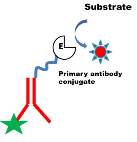 Figure 1. Diagrammatic representation of detection of antigen in direct ELISA. (Malhotra et al., 2025)
Signal Generation Strategy
Signal system optimization begins with rational pairing of the enzyme, substrate, reaction environment, and cofactor requirements. We treat these elements as an integrated system rather than isolated reagents.
Enzyme Selection
- Horseradish peroxidase (HRP) selected for rapid-turnover, high-signal applications where burst kinetics and broad substrate compatibility are advantageous; optimal for chromogenic and chemiluminescent readouts in ELISA, Western blot, and IHC platforms → HRP Conjugation Service for Immunoassays
- Alkaline phosphatase (AP) chosen for assays requiring sustained signal accumulation, broad dynamic range, and low endogenous background; preferred for quantitative ELISA, CLIA, and applications where extended incubation improves low-end discrimination → Alkaline Phosphatase (AP) Conjugation Service
- Specialty enzymes (β-galactosidase, urease, glucose oxidase, luciferase) deployed for ultra-sensitive fluorescence, electrochemical pH-shift, or coupled amplification cascades where standard reporters fail to meet sensitivity or instrumentation constraints → β-Galactosidase, Urease and Oxidase Labeling Services
- Enzyme source and isoform evaluated for specific activity, glycosylation profile, thermal stability, and compatibility with the intended assay temperature and incubation time
Substrate Screening
- Chromogenic substrates (TMB, ABTS, OPD for HRP; pNPP for AP; ONPG for β-galactosidase) screened for molar extinction coefficient, solubility, and kinetic turnover rate to match the required sensitivity and endpoint stability
- Chemiluminescent substrates (luminol/enhancer systems, acridan esters for HRP; CSPD, CDP-Star for AP; Galacton-Star for β-galactosidase) evaluated for glow kinetics, signal half-life, and compatibility with luminometer integration times
- Fluorogenic substrates (Amplex Red, H2DCFDA for HRP-coupled systems; 4-MUP for AP; MUG, resorufin-β-galactoside for β-galactosidase) assessed for quantum yield, Stokes shift, and freedom from assay component autofluorescence
- Substrate solubility and precipitation potential tested under assay conditions to prevent turbidity interference and microplate reader artifacts
Buffer Optimization
- pH titration across the enzyme's active range to identify the optimum that maximizes catalytic rate while preserving conjugate stability; for HRP, pH 5.0–6.5 optimizes compound I formation, whereas AP requires pH 9.0–10.5 for maximal pNPP turnover
- Ionic strength optimization (NaCl, KCl concentration) to balance enzyme activity against non-specific binding and electrostatic aggregation; high salt can suppress HRP activity while improving AP signal-to-background in serum matrices
- Buffer species selection (phosphate, Tris, MOPS, HEPES, carbonate, diethanolamine) evaluated for metal chelation, enzyme inhibition, and compatibility with substrate solubility; phosphate buffers avoided for AP assays due to product inhibition by inorganic phosphate
- Surfactant and blocking agent screening (Tween-20, Triton X-100, BSA, casein, fish gelatin) to minimize non-specific signal without inhibiting enzyme activity or inducing substrate micelle formation
Cofactor Optimization
- AP cofactors: Zn2+ and Mg2+ supplementation (0.1–1.0 mM) verified as essential for maintaining the binuclear metal center and allosteric activation; EDTA-free buffers mandated to prevent apoenzyme formation
- β-galactosidase cofactors: Mg2+ and K+ included to stabilize the tetrameric quaternary structure and enhance catalytic turnover; Na+ and Li+ evaluated as alternative monovalent activators
- Oxidase cofactors: FAD retention in glucose oxidase and related flavoproteins optimized through pH control (pH 5.5–7.0) and avoidance of chaotropic agents that promote flavin dissociation
- HRP enhancers: Phenolic enhancers (4-iodophenol, 4-hydroxycinnamic acid) and chemiluminescent enhancers (firefly luciferin analogs) titrated to amplify signal output without increasing background oxidation
Signal Performance Optimization
Beyond reagent selection, we engineer the reaction conditions and assay architecture to maximize sensitivity, linearity, background suppression, and kinetic control.
Sensitivity
- Substrate concentration titrated to ensure zero-order kinetics ([S] >> KM) during the assay window, maximizing the rate of product formation and lowering the limit of detection (LOD) by extending the signal-to-noise ratio at low analyte concentrations
- Enzyme conjugate working concentration optimized to the lowest dilution that still yields robust signal, reducing reagent consumption and minimizing hook effects in sandwich assays
- Signal amplification strategies evaluated, including poly-HRP polymers, HRP-AP coupled cascade systems, and tyramide signal amplification (TSA) for applications requiring sub-picomolar detection limits
Dynamic Range
- Substrate depletion management through controlled reaction time, substrate replenishment protocols, or stop-solution chemistry to prevent signal saturation at high analyte concentrations and extend the upper limit of quantification (ULOQ)
- AP-based systems leveraged for their inherently broader linear range compared to HRP burst kinetics, enabling three- to four-log quantification without sample dilution and re-assay
- Standard curve modeling (4-parameter logistic, 5-parameter logistic, spline fitting) optimized for the specific sigmoidal characteristics of each enzyme-substrate system to improve accuracy at curve extremes
Background Suppression
- Substrate blank optimization to identify and eliminate lot-to-lot variation in substrate auto-oxidation or intrinsic absorbance; high-purity substrate lots qualified against acceptance criteria for baseline optical density
- H2O2 contamination control in HRP systems through catalase pre-treatment of buffers, sealed reagent packaging, and stabilizer selection (e.g., glycerol, non-reactive polyols) that suppress non-enzymatic peroxide generation
- Endogenous enzyme quenching protocols (levamisole for AP, sodium azide for HRP) validated for compatibility with the reporter enzyme while effectively suppressing sample-derived background in tissue, serum, and environmental matrices
Reaction Kinetics
- Real-time kinetic profiling to determine the linear phase of product accumulation, establishing the optimal readout window that maximizes signal while remaining within the initial-rate regime for quantitative accuracy
- Temperature optimization (25°C, 37°C, room temperature) to balance reaction velocity against reagent stability and assay workflow constraints; Arrhenius analysis applied to predict rate constants at non-standard temperatures
- Stop-solution chemistry (sulfuric acid for TMB, EDTA for metal-dependent enzymes, heat denaturation) validated to arrest reaction kinetics instantaneously and uniformly across the microplate, ensuring inter-well consistency in endpoint assays
Signal Detection Systems
The choice of detection modality fundamentally constrains the achievable sensitivity and throughput. We optimize the enzyme-substrate system for seamless integration with the client's instrumentation.
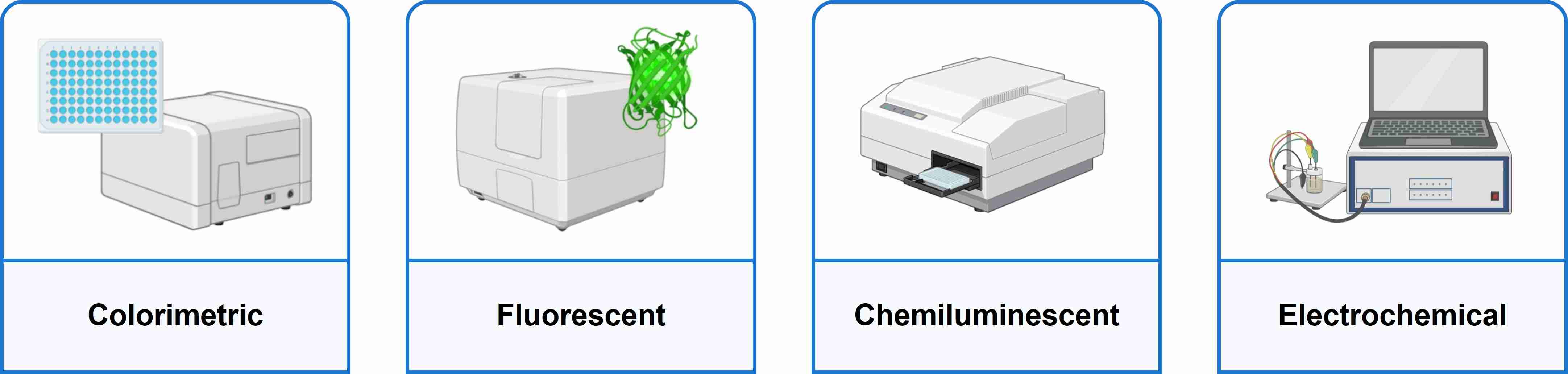
Colorimetric
- Absorbance-based detection at enzyme-specific wavelengths (450 nm for TMB; 405 nm for pNPP; 490 nm for OPD) with pathlength correction and reference wavelength subtraction (620–650 nm) to compensate for meniscus effects and microplate imperfections
- Microplate reader linearity validation (0–3.0 OD) to ensure that high-signal wells remain within the instrument's dynamic range; substrate concentrations adjusted to prevent absorbance saturation in strongly positive samples
- Compatibility with standard ELISA readers and spectrophotometers without specialized equipment, making colorimetric systems ideal for resource-limited settings and high-throughput screening
Fluorescent
- Fluorogenic substrate selection based on excitation/emission spectra compatibility with the instrument's filter sets or monochromator range; 4-MUP (Ex/Em 360/440 nm), resorufin (Ex/Em 570/585 nm), and coumarin derivatives evaluated for minimal spectral overlap in multiplexed panels
- Autofluorescence mitigation through black microplates, sample matrix blank subtraction, and buffer formulation free of fluorescent contaminants (aromatic preservatives, certain detergents)
- Signal stability under excitation light exposure assessed to identify substrates susceptible to photobleaching; read protocols optimized to minimize exposure time while maintaining signal-to-noise integrity
Chemiluminescent
- Glow-type vs. flash-type substrate selection matched to reader capabilities: glow substrates (enhanced luminol, dioxetanes) provide stable light emission over 5–60 minutes, compatible with batch reading and automated platforms; flash substrates require injectors and immediate capture
- Signal integration time optimization to maximize relative light units (RLU) while minimizing well-to-well variability; typical integration windows of 0.1–1.0 seconds per well for standard luminometers
- Cross-talk and plate edge effects mitigated through opaque white microplates, optimized reagent volume, and spatial calibration to ensure uniform light collection across the entire plate surface
Electrochemical
- Amperometric detection of H2O2 generated by oxidase-coupled or direct oxidase systems at platinum, carbon nanotube, or screen-printed carbon electrodes; applied potential optimized (typically +0.6 to +0.7 V vs. Ag/AgCl) to maximize Faradaic current while minimizing interferent oxidation
- Oxygen depletion monitoring via Clark-type electrodes for oxidase-based biosensors, with calibration curves establishing the linear relationship between analyte concentration and current change
- pH-shift detection using ion-selective field-effect transistors (ISFETs) or iridium oxide electrodes for urease-coupled systems, enabling label-free, reagent-less detection architectures suitable for continuous monitoring and wearable diagnostics
Typical Applications
Our signal system optimization service is applied across the major diagnostic platforms where enzyme-generated signal quality directly determines clinical utility.
- ELISA: Optimization of TMB, ABTS, and pNPP endpoint systems for sandwich, competitive, and indirect ELISA formats. We tune substrate concentration, reaction time, and stop chemistry to maximize signal-to-background ratios and extend dynamic range across cytokine, hormone, infectious disease, and autoantibody panels. Automated liquid handler compatibility and plate reader calibration support are included.
- CLIA: Chemiluminescence substrate optimization for automated analyzer platforms, including enhanced luminol systems for HRP and dioxetane substrates (CSPD, CDP-Star) for AP. We validate glow kinetics, signal stability during batch reading, and substrate lot consistency to meet the stringent precision requirements (CV
- Biosensors: Electrochemical signal system design for amperometric glucose, lactate, and cholesterol biosensors, including H2O2 detection optimization, mediator selection (ferrocene derivatives, Prussian blue), and electrode surface modification to enhance electron transfer kinetics and operational stability.
- POCT: Signal system miniaturization for lateral flow, microfluidic, and cartridge-based point-of-care devices. We develop dry-reagent formulations with rapid rehydration kinetics, ambient-temperature stability, and visual or smartphone-readable colorimetric endpoints that eliminate the need for dedicated instrumentation in resource-limited settings.
FAQs
Q1. How much can signal system optimization improve my assay sensitivity?
A1. Systematic optimization of the enzyme-substrate pair, buffer matrix, and detection conditions typically improves LOD by 2- to 10-fold compared to off-the-shelf reagent protocols. In cases where we implement amplification cascades (poly-HRP, TSA, or coupled enzyme systems), sensitivity improvements of 50- to 100-fold are achievable. The exact gain depends on the starting assay architecture and the analyte concentration range targeted.
Q2. Will changing the substrate affect my assay's regulatory validation?
A2. Any substrate change in a regulated diagnostic assay requires re-validation of analytical performance (precision, accuracy, linearity, LOD, interference studies) under the updated protocol. We design optimization campaigns with regulatory compliance in mind, generating the full validation data package—including stability studies, reference range verification, and lot-to-lot consistency data—required for 510(k), IVDR, or CLIA-waived submission support.
Q3. Can you optimize signal systems for enzymes we already manufacture in-house?
A3. Yes. We routinely optimize substrate formulations, buffer systems, and detection protocols for client-produced enzymes. Our service is agnostic to enzyme source—we work with your existing HRP, AP, or specialty enzyme lots, characterizing their specific activity profiles and designing signal systems that extract maximum performance from your proprietary reagents.
Q4. What is the difference between glow and flash chemiluminescence, and which should I choose?
A4. Flash chemiluminescence produces a rapid signal burst (peak within seconds) that requires immediate detection, typically with injector-equipped luminometers. Glow chemiluminescence generates sustained light emission over minutes to hours, enabling batch reading on standard plate readers without injectors. Choose flash for maximum peak intensity and lowest detection limits; choose glow for workflow flexibility, automated platform compatibility, and reduced instrument complexity.
Q5. How do you handle substrate lot-to-lot variability?
A5. We qualify every substrate lot against a master reference standard, measuring auto-oxidation rate, purity by HPLC, and performance in a standardized enzyme activity assay. Lots failing to meet specification (typically ±5% of reference activity) are rejected. For regulated manufacturing, we establish substrate release criteria and vendor qualification programs to ensure long-term consistency.
Q6. Can you develop signal systems for multiplexed assays with multiple enzymes?
A6. Yes. We design orthogonal signal systems for multiplexed detection, pairing enzymes with spectrally distinct substrates (e.g., HRP-TMB colorimetric with AP-fluorogenic, or dual chemiluminescent substrates with non-overlapping emission kinetics). We also engineer spatially resolved detection on microarray and bead-based platforms, ensuring that signal from one enzyme channel does not cross-interfere with another.
Creative Enzymes Diagnostic approaches signal generation as a systems engineering challenge, integrating enzyme biochemistry, substrate kinetics, buffer formulation science, and detection physics into a unified optimization workflow. By looking beyond the conjugate to the complete signal cascade, we deliver assay systems with superior sensitivity, broader dynamic range, and robust reproducibility—positioning your diagnostic platform ahead of competitors who treat signal chemistry as an afterthought.
Contact our business development team today to discuss your signal system optimization requirements!
References
- Malhotra N, Chahal A, Jain A, et al. Monoclonal antibodies: purification, application in conventional methods and cutting edge technology. Biomedical Materials & Devices. 2025;3(1):367-380. doi:10.1007/s44174-024-00203-2
Related Services

