
Companion diagnostic assays often need to detect biomarkers present at low abundance against complex biological background. Signal amplification makes this possible by increasing the measurable output associated with a target. The amplification may occur at the target level, through repeated copying of nucleic acid; at the reporter level, through catalytic turnover; or through a layered cascade that recruits multiple labels or reactions. Each strategy improves sensitivity in a different way and introduces its own sources of bias, background, saturation, and variability.
This article compares major amplification approaches used in molecular, immunoassay, and biosensor CDx workflows. It emphasizes design tradeoffs and analytical controls rather than treating maximum signal as the goal.
Amplification increases the difference between a target-associated event and the baseline response. In a nucleic acid assay, the target itself or a target-derived sequence may be copied. In an immunoassay, a reporter enzyme can generate many signal molecules after one binding event. Other systems use branched probes, nanoparticles, deposition chemistries, or coupled enzyme reactions to multiply labels or outputs.
Amplification is useful only when it improves decision quality. A stronger signal is not necessarily a better assay. If background, variability, or saturation rises at the same time, the clinically relevant signal-to-noise ratio or classification agreement may not improve.
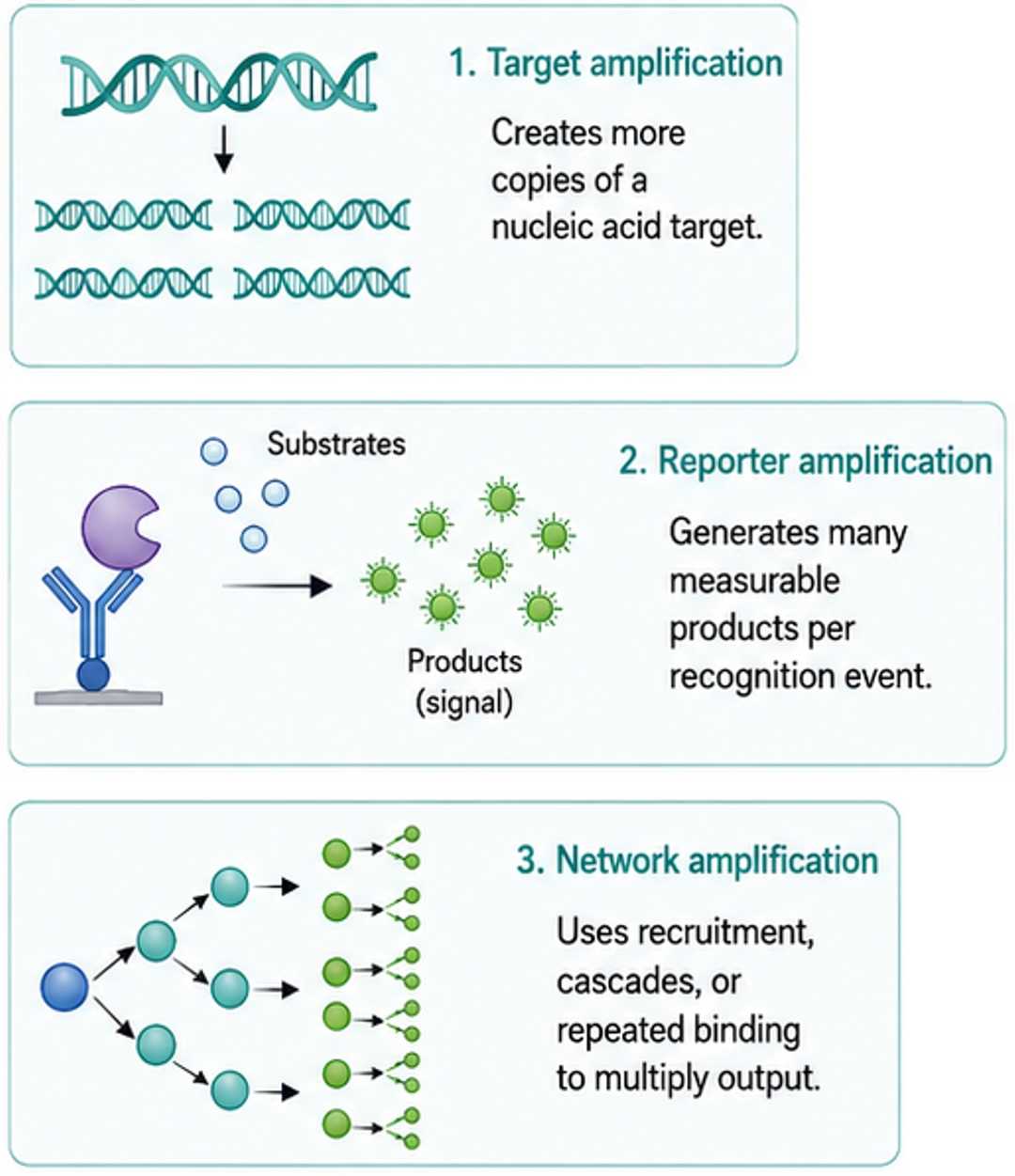
Figure 1. Amplification can occur at different layers of a CDx measurement system.
PCR uses repeated thermal cycles to copy a defined nucleic acid region. In qPCR, fluorescence is measured during amplification, allowing the accumulation curve to support qualitative or quantitative interpretation. Hot-start polymerases can reduce extension before cycling begins, while probe-based chemistries add sequence-specific recognition. Multiplex formats measure several targets or controls in one reaction but require careful balancing of primers, probes, enzyme capacity, and fluorescence channels.
For CDx applications, sensitivity must be evaluated alongside allele discrimination and error control. Increasing cycle number can reveal lower inputs, but it can also expose low-level contamination and nonspecific products. Low-frequency variant detection requires a defined limit of blank, limit of detection, and reproducibility at relevant allele fractions. A theoretical copy calculation does not replace empirical detection probability across runs, lots, operators, and specimen backgrounds.
Established workflows, flexible primer design, broad instrument availability, and quantitative real-time readout.
Primer mismatch, inhibition, contamination, differential efficiency, and competition in multiplex reactions.
Extraction control, internal amplification control, no-template control, and near-cutoff positive material.
Representative components include Taq HS DNA Polymerase and qPCR Probe Master Mix. Developers needing assay-specific balancing can use PCR and qPCR enzyme/premix development.
Isothermal methods amplify nucleic acids at a relatively constant temperature and can reduce instrument complexity. LAMP uses multiple primers and a strand-displacing polymerase to generate large quantities of product. Other isothermal architectures use recombinases, helicases, nicking enzymes, or transcription-based cycles. Their speed and equipment-light operation can be attractive for point-of-care settings, but reaction behavior is often highly dependent on primer design, temperature, and contamination control.
A rapid visual endpoint is convenient, yet color can be affected by buffering capacity, specimen chemistry, or nonspecific amplification. Fluorescence provides kinetic information but still requires threshold and timing rules. Closed-tube workflows help contain high-copy products. Lyophilized formats may simplify distribution, but drying and reconstitution can alter enzyme recovery and reaction balance.
| Design question | Why it matters | Practical experiment |
|---|---|---|
| How quickly should positives appear? | Late nonspecific reactions can approach the decision window | Test time-to-positive distributions for negatives and low positives |
| How stable is temperature? | Small shifts may change enzyme and primer kinetics | Challenge the allowed heater and ambient range |
| Can matrix chemistry change readout? | pH or optical effects can mimic a result | Evaluate representative specimens and interferents |
| Will dried reagents recover uniformly? | Incomplete dissolution creates reaction-to-reaction variation | Study wetting, mixing, recovery time and fill position |
Examples include ColorDetect LAMP/RT-LAMP Mix and the Fluorescent LAMP/RT-LAMP Kit. The LAMP and RT-LAMP reagent development service supports format-specific optimization.
In enzyme-linked detection, an antibody, probe, or other recognition element carries an enzyme label. Horseradish peroxidase, alkaline phosphatase, beta-galactosidase, and oxidases can convert many substrate molecules after one target-binding event. Colorimetric systems are simple and widely compatible; fluorescent and chemiluminescent substrates can provide greater sensitivity; electrochemical products can suit compact biosensors.
The conjugate is a coupled reagent. Label density affects catalytic capacity, antibody affinity, aggregation, and transport through porous materials. Substrate concentration, reaction time, mixing, temperature, and stop chemistry affect the measured result. When enzyme turnover is fast, small timing differences may become large signal differences. A slower, more stable reaction can sometimes provide better precision and a wider operational window.
Background deserves equal priority. Nonspecific conjugate retention, endogenous enzyme activity, substrate autoxidation, inadequate washing, and surface adsorption can all be amplified. Blank signal, low-positive separation, and high-dose behavior should be considered together. Enzyme–antibody conjugates, custom conjugate optimization, and enzyme–substrate system optimization are relevant resources for this design space.
Coupled reactions use the product of one enzyme as the substrate or cofactor input for another reaction. They can convert an analyte that is difficult to measure directly into a convenient optical or electrochemical output. Cascades may also regenerate cofactors or repeatedly cycle a reporter species. In immunodetection, secondary antibodies, avidin–biotin systems, polymeric labels, and deposition reactions increase the number of reporters associated with a target.
Cascade design requires kinetic matching. If the first reaction is rate-limiting, increasing the reporter enzyme may not improve sensitivity. If the downstream reaction is too fast, it may saturate before meaningful differences are resolved. The substrate, cofactor, and coupled reaction design service addresses this system-level balance.
| Priority | Potentially suitable strategy | Tradeoff to examine |
|---|---|---|
| Very low-copy DNA target | qPCR, digital PCR, or targeted preamplification | Contamination and introduced error |
| Rapid equipment-light nucleic acid result | Isothermal amplification | Primer complexity and late nonspecific signal |
| Low-abundance protein | Enzyme reporter or layered immunoassay | Nonspecific binding and conjugate variability |
| Small metabolite or enzyme activity | Coupled catalytic cascade | Matrix effects and kinetic coupling |
| Multiplex molecular profile | Multiplex PCR or NGS library amplification | Competition, bias, coverage, and algorithm control |
Selection begins with the intended clinical decision, not with the most sensitive available chemistry. Developers should define target prevalence, specimen input, acceptable time to result, instrument constraints, throughput, multiplex needs, and consequences of false results. A strategy that works in purified buffer may fail in tissue extracts or blood-derived material. A strategy that reaches an impressive analytical limit may have poor reproducibility at the decision point.
Optimize separation, not brightness. The useful target is stable discrimination between clinically relevant groups with controlled invalid rates, not the highest possible raw signal.
Every amplification method preferentially magnifies something. Ideally that is the intended target; in practice, it can also magnify contamination, off-target priming, nonspecific binding, optical background, or minor handling differences. Controls should localize the failure. A process control can reveal extraction loss, an internal amplification control can reveal inhibition, and a blank can reveal contamination or reporter background. Positive controls near the claimed detection limit challenge the most vulnerable range.
Dynamic range must be protected at both ends. At low signal, stochastic sampling can dominate. At high signal, reagent depletion, detector saturation, hook effects, or plateau amplification can compress differences. Dilution, alternate calibration, or a second measurement range may be preferable to forcing one amplification setting to cover all samples. Robustness studies should vary temperature, timing, reagent volume, operator, lot, and instrument within realistic limits.
Data analysis is part of the system. Baseline subtraction, fluorescence thresholds, image exposure, curve-fitting, and classification algorithms must be controlled. Changing the algorithm after wet-lab validation can change clinical calls even if the chemistry is unchanged. Limit-of-detection optimization should therefore evaluate the end-to-end measurement process.
Transfer should include raw data examples, curve interpretation, control trends, and troubleshooting criteria. An amplification reaction that depends on tacit timing or manual judgment is difficult to reproduce. CDx assay transfer and manufacturability assessment can reveal these hidden dependencies before scale-up.
Before design lock, reviewers should be able to explain what is amplified, how amplification terminates or is read, and which nonspecific events receive the same gain. They should identify the useful measurement window, the first source of saturation, and the variables that most strongly change time to result or endpoint intensity. Control materials should demonstrate extraction, reaction, and analysis performance without creating an unrealistic signal margin.
The review should also examine deployment. A chemistry that is reliable with automated timing may be fragile in a manual workflow. A highly sensitive open-tube method may create carryover risk at scale. A multistep cascade may be difficult to dry or transfer. These are not reasons to reject amplification; they determine the controls, packaging, automation, training, and acceptance criteria needed to use it responsibly in a CDx assay.
Signal amplification enables CDx assays to measure low-abundance biomarkers, but gain must be balanced against background, bias, saturation, and operational variability. PCR, isothermal amplification, enzyme reporters, and coupled cascades amplify different parts of the measurement chain. The best strategy is the one that produces reproducible separation at the clinically relevant decision point and remains controllable across specimens, reagent lots, instruments, sites, and storage conditions.
We support CDx signal development through enzyme and master-mix selection, conjugation, coupled-reaction design, background reduction, sensitivity studies, and assay transfer.
